 Movie
Movie Controller

[English] 日本語
 Yorodumi
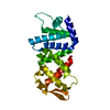
- PDB-1t8b: Crystal structure of refolded PHOU-like protein (gi 2983430) from... -
+ Open data

- Basic information
| Entry | Database: PDB / ID: 1t8b | ||||||
|---|---|---|---|---|---|---|---|
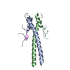
| Title | Crystal structure of refolded PHOU-like protein (gi 2983430) from Aquifex aeolicus | ||||||
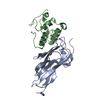
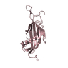
 Components Components | Phosphate transport system protein phoU homolog | ||||||
 Keywords Keywords | TRANSPORT PROTEIN / alpha-helical protein consisting of two 3-helix bundles / Structural Genomics / BSGC structure funded by NIH / Protein Structure Initiative / PSI / Berkeley Structural Genomics Center | ||||||
| Function / homology |  Function and homology information Function and homology informationnegative regulation of phosphate transmembrane transport / negative regulation of phosphate metabolic process / phosphate ion transport / intracellular phosphate ion homeostasis / negative regulation of gene expression / protein homodimerization activity / cytoplasm Similarity search - Function | ||||||
| Biological species |   Aquifex aeolicus (bacteria) Aquifex aeolicus (bacteria) | ||||||
| Method |  X-RAY DIFFRACTION / SYNCHROTRON / SAD, molecular replacement / Resolution: 3.23 Å X-RAY DIFFRACTION / SYNCHROTRON / SAD, molecular replacement / Resolution: 3.23 Å | ||||||
 Authors Authors | Oganesyan, V. / Kim, S.-H. / Oganesyan, N. / Jancarik, J. / Adams, P.D. / Kim, R. / Berkeley Structural Genomics Center (BSGC) | ||||||
 Citation Citation | Journal: J.Bacteriol. / Year: 2005 Title: Crystal structure of the "PhoU-like" phosphate uptake regulator from Aquifex aeolicus. Authors: Oganesyan, V. / Oganesyan, N. / Adams, P.D. / Jancarik, J. / Yokota, H.A. / Kim, R. / Kim, S.H. | ||||||
| History |
|
- Structure visualization
| Structure viewer | Molecule: MolmilJmol/JSmol |
|---|
- Downloads & links
-Download
| PDBx/mmCIF format | 1t8b.cif.gz | 91.7 KB | Display | PDBx/mmCIF format |
|---|---|---|---|---|
| PDB format | pdb1t8b.ent.gz | 71.3 KB | Display | PDB format |
| PDBx/mmJSON format | 1t8b.json.gz | Tree view | PDBx/mmJSON format | |
| Others |  Other downloads Other downloads |
-Validation report
| Arichive directory | https://data.pdbj.org/pub/pdb/validation_reports/t8/1t8bftp://data.pdbj.org/pub/pdb/validation_reports/t8/1t8b | HTTPS FTP |
|---|
-Related structure data
| Related structure data | 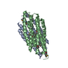 1t72SC S: Starting model for refinement C: citing same article ( |
|---|---|
| Similar structure data | |
| Other databases |
-Links
 PDBj
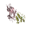
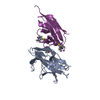
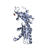
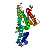
PDBj- Assembly
| Deposited unit | 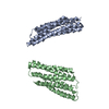
| ||||||||||||||||||
|---|---|---|---|---|---|---|---|---|---|---|---|---|---|---|---|---|---|---|---|
| 1 | 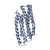
| ||||||||||||||||||
| 2 | 
| ||||||||||||||||||
| 3 | 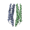
| ||||||||||||||||||
| Unit cell |
| ||||||||||||||||||
| Noncrystallographic symmetry (NCS) | NCS domain:
NCS domain segments: Component-ID: 1 / Ens-ID: 1 / Beg auth comp-ID: MET / Beg label comp-ID: MET / End auth comp-ID: GLY / End label comp-ID: GLY / Refine code: _ / Auth seq-ID: 7 - 214 / Label seq-ID: 7 - 214
| ||||||||||||||||||
| Details | Biological assembly is monomeric |
-Components
| #1: Protein | Mass: 25944.762 Da / Num. of mol.: 2 Source method: isolated from a genetically manipulated source Source: (gene. exp.) Aquifex aeolicus (bacteria) / Gene: PHOU, AQ_906 / Plasmid: pB4.1105B / Species (production host): Escherichia coli / Production host: |
|---|
-Experimental details
-Experiment
| Experiment | Method: X-RAY DIFFRACTION / Number of used crystals: 1 |
|---|
- Sample preparation
| Crystal | Density Matthews: 2.5 Å3/Da / Density % sol: 51 % |
|---|---|
| Crystal grow | Temperature: 298 K / Method: vapor diffusion, hanging drop / pH: 7.5 Details: Lithium Sulfate, pH 7.5, VAPOR DIFFUSION, HANGING DROP, temperature 298K |
-Data collection
| Diffraction | Mean temperature: 100 K |
|---|---|
| Diffraction source | Source: SYNCHROTRON / Site: ALS  / Beamline: 5.0.2 / Wavelength: 0.9792 Å / Beamline: 5.0.2 / Wavelength: 0.9792 Å |
| Detector | Type: ADSC QUANTUM 4 / Detector: CCD / Date: Jan 10, 2004 / Details: double Si(111) crystal |
| Radiation | Monochromator: double Si(111) crystal / Protocol: SINGLE WAVELENGTH / Monochromatic (M) / Laue (L): M / Scattering type: x-ray |
| Radiation wavelength | Wavelength: 0.9792 Å / Relative weight: 1 |
| Reflection | Resolution: 3.23→48 Å / Num. obs: 8109 / % possible obs: 99.3 % / Observed criterion σ(F): 0 / Observed criterion σ(I): 0 / Redundancy: 12.2 % / Biso Wilson estimate: 120 Å2 / Rmerge(I) obs: 0.109 / Rsym value: 0.109 / Net I/σ(I): 14 |
| Reflection shell | Resolution: 3.23→3.393 Å / Redundancy: 2 % / Rmerge(I) obs: 0.78 / Mean I/σ(I) obs: 2.1 / Rsym value: 0.78 / % possible all: 90 |
- Processing
| Software |
| ||||||||||||||||||||||||||||||||||||||||||||||||||||||||||||||||||||||||||||||||||||||||||||||||||||||||||||||||||||||||||||||||||||||||||||||||||||||||||||||||||||||||||
|---|---|---|---|---|---|---|---|---|---|---|---|---|---|---|---|---|---|---|---|---|---|---|---|---|---|---|---|---|---|---|---|---|---|---|---|---|---|---|---|---|---|---|---|---|---|---|---|---|---|---|---|---|---|---|---|---|---|---|---|---|---|---|---|---|---|---|---|---|---|---|---|---|---|---|---|---|---|---|---|---|---|---|---|---|---|---|---|---|---|---|---|---|---|---|---|---|---|---|---|---|---|---|---|---|---|---|---|---|---|---|---|---|---|---|---|---|---|---|---|---|---|---|---|---|---|---|---|---|---|---|---|---|---|---|---|---|---|---|---|---|---|---|---|---|---|---|---|---|---|---|---|---|---|---|---|---|---|---|---|---|---|---|---|---|---|---|---|---|---|---|---|
| Refinement | Method to determine structure: SAD, molecular replacement Starting model: pdb entry 1T72 Resolution: 3.23→15 Å / Cor.coef. Fo:Fc: 0.96 / Cor.coef. Fo:Fc free: 0.94 / SU B: 33.07 / SU ML: 0.532 / TLS residual ADP flag: LIKELY RESIDUAL / Isotropic thermal model: Isotropic / Cross valid method: THROUGHOUT / σ(F): 1 / ESU R Free: 0.541 / Stereochemistry target values: MAXIMUM LIKELIHOOD / Details: Matrix scaling, diagonal term 0.02
| ||||||||||||||||||||||||||||||||||||||||||||||||||||||||||||||||||||||||||||||||||||||||||||||||||||||||||||||||||||||||||||||||||||||||||||||||||||||||||||||||||||||||||
| Solvent computation | Ion probe radii: 0.8 Å / Shrinkage radii: 0.8 Å / VDW probe radii: 1.4 Å / Solvent model: BABINET MODEL WITH MASK | ||||||||||||||||||||||||||||||||||||||||||||||||||||||||||||||||||||||||||||||||||||||||||||||||||||||||||||||||||||||||||||||||||||||||||||||||||||||||||||||||||||||||||
| Displacement parameters | Biso mean: 128.948 Å2
| ||||||||||||||||||||||||||||||||||||||||||||||||||||||||||||||||||||||||||||||||||||||||||||||||||||||||||||||||||||||||||||||||||||||||||||||||||||||||||||||||||||||||||
| Refine analyze |
| ||||||||||||||||||||||||||||||||||||||||||||||||||||||||||||||||||||||||||||||||||||||||||||||||||||||||||||||||||||||||||||||||||||||||||||||||||||||||||||||||||||||||||
| Refinement step | Cycle: LAST / Resolution: 3.23→15 Å
| ||||||||||||||||||||||||||||||||||||||||||||||||||||||||||||||||||||||||||||||||||||||||||||||||||||||||||||||||||||||||||||||||||||||||||||||||||||||||||||||||||||||||||
| Refine LS restraints |
| ||||||||||||||||||||||||||||||||||||||||||||||||||||||||||||||||||||||||||||||||||||||||||||||||||||||||||||||||||||||||||||||||||||||||||||||||||||||||||||||||||||||||||
| Refine LS restraints NCS | Dom-ID: 1 / Auth asym-ID: A / Ens-ID: 1 / Number: 1679 / Refine-ID: X-RAY DIFFRACTION
| ||||||||||||||||||||||||||||||||||||||||||||||||||||||||||||||||||||||||||||||||||||||||||||||||||||||||||||||||||||||||||||||||||||||||||||||||||||||||||||||||||||||||||
| LS refinement shell | Resolution: 3.23→3.393 Å / Total num. of bins used: 10 /
| ||||||||||||||||||||||||||||||||||||||||||||||||||||||||||||||||||||||||||||||||||||||||||||||||||||||||||||||||||||||||||||||||||||||||||||||||||||||||||||||||||||||||||
| Refinement TLS params. | Method: refined / Refine-ID: X-RAY DIFFRACTION
| ||||||||||||||||||||||||||||||||||||||||||||||||||||||||||||||||||||||||||||||||||||||||||||||||||||||||||||||||||||||||||||||||||||||||||||||||||||||||||||||||||||||||||
| Refinement TLS group |
|
