 Movie
Movie Controller


+ Open data

- Basic information
| Entry | Database: PDB / ID: 1t4i | ||||||
|---|---|---|---|---|---|---|---|
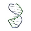
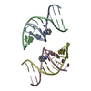
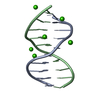
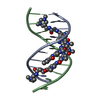
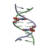
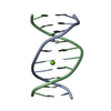
| Title | Crystal Structure of a DNA Decamer Containing a Thymine-dimer | ||||||
 Components Components |
| ||||||
 Keywords Keywords | DNA / UV-damaged DNA / Thymine dimer / DNA decamer | ||||||
| Function / homology | DNA Function and homology information Function and homology information | ||||||
| Method |  X-RAY DIFFRACTION / MOLECULAR REPLACEMENT / Resolution: 2.5 Å X-RAY DIFFRACTION / MOLECULAR REPLACEMENT / Resolution: 2.5 Å | ||||||
 Authors Authors | Park, H. / Zhang, K. / Ren, Y. / Nadji, S. / Sinha, N. / Taylor, J.S. / Kang, C. | ||||||
 Citation Citation | Journal: Proc.Natl.Acad.Sci.USA / Year: 2002 Title: Crystal Structure of a DNA Decamer Containing a cis-syn Thymine-dimer Authors: Park, H. / Zhang, K. / Ren, Y. / Nadji, S. / Sinha, N. / Taylor, J.S. / Kang, C. | ||||||
| History |
|
- Structure visualization
| Structure viewer | Molecule: MolmilJmol/JSmol |
|---|
- Downloads & links
-Download
| PDBx/mmCIF format | 1t4i.cif.gz | 34.4 KB | Display | PDBx/mmCIF format |
|---|---|---|---|---|
| PDB format | pdb1t4i.ent.gz | 21.6 KB | Display | PDB format |
| PDBx/mmJSON format | 1t4i.json.gz | Tree view | PDBx/mmJSON format | |
| Others |  Other downloads Other downloads |
-Validation report
| Arichive directory | https://data.pdbj.org/pub/pdb/validation_reports/t4/1t4iftp://data.pdbj.org/pub/pdb/validation_reports/t4/1t4i | HTTPS FTP |
|---|
-Related structure data


-Links
 PDBj
PDBj


- Assembly
| Deposited unit | 
| ||||||||
|---|---|---|---|---|---|---|---|---|---|
| 1 | 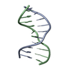
| ||||||||
| 2 | 
| ||||||||
| Unit cell |
|
-Components
| #1: DNA chain | Mass: 3035.003 Da / Num. of mol.: 2 / Source method: obtained synthetically #2: DNA chain | Mass: 3053.031 Da / Num. of mol.: 2 / Source method: obtained synthetically #3: Water | ChemComp-HOH / |  Mass: 18.015 Da / Num. of mol.: 56 / Source method: isolated from a natural source / Formula: H2O Mass: 18.015 Da / Num. of mol.: 56 / Source method: isolated from a natural source / Formula: H2O |
|---|
-Experimental details
-Experiment
| Experiment | Method: X-RAY DIFFRACTION / Number of used crystals: 1 |
|---|
- Sample preparation
| Crystal grow | Temperature: 277 K / Method: vapor diffusion, hanging drop / pH: 7.5 Details: MgCl2, HEPES-Na, PEG 400, pH 7.5, VAPOR DIFFUSION, HANGING DROP, temperature 277K | ||||||||||||||||||||||||||||||||
|---|---|---|---|---|---|---|---|---|---|---|---|---|---|---|---|---|---|---|---|---|---|---|---|---|---|---|---|---|---|---|---|---|---|
| Components of the solutions |
|
-Data collection
| Diffraction | Mean temperature: 100 K |
|---|---|
| Diffraction source | Source: ROTATING ANODE / Type: RIGAKU / Wavelength: 1.5418 Å |
| Detector | Type: RIGAKU RAXIS IIC / Detector: IMAGE PLATE / Date: Jan 1, 1999 / Details: mirror |
| Radiation | Monochromator: Ni Filter / Protocol: SINGLE WAVELENGTH / Monochromatic (M) / Laue (L): M / Scattering type: x-ray |
| Radiation wavelength | Wavelength: 1.5418 Å / Relative weight: 1 |
| Reflection | Resolution: 2.3→20 Å / Num. all: 5775 / Num. obs: 4616 / % possible obs: 88.6 % / Observed criterion σ(F): 1 / Observed criterion σ(I): 1 |
| Reflection shell | Resolution: 2.3→2.39 Å / % possible all: 76.4 |
- Processing
| Software |
| ||||||||||||||||||||
|---|---|---|---|---|---|---|---|---|---|---|---|---|---|---|---|---|---|---|---|---|---|
| Refinement | Method to determine structure: MOLECULAR REPLACEMENT / Resolution: 2.5→10 Å / σ(F): 2.3
| ||||||||||||||||||||
| Refinement step | Cycle: LAST / Resolution: 2.5→10 Å
| ||||||||||||||||||||
| LS refinement shell | Resolution: 2.5→2.61 Å /
|
