 Movie
Movie Controller

[English] 日本語
 Yorodumi
- PDB-1t2q: The Crystal Structure of an NNA7 Fab that recognizes an N-type bl... -
+ Open data

- Basic information
| Entry | Database: PDB / ID: 1t2q | ||||||
|---|---|---|---|---|---|---|---|
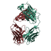
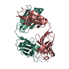
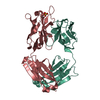
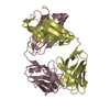
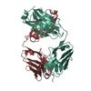
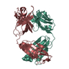
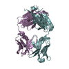
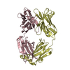
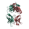
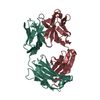
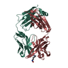
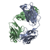
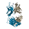
| Title | The Crystal Structure of an NNA7 Fab that recognizes an N-type blood group antigen | ||||||
 Components Components |
| ||||||
 Keywords Keywords | IMMUNE SYSTEM / Fab / glycophorin A / blood group antigen | ||||||
| Function / homology |  Function and homology information Function and homology informationimmunoglobulin mediated immune response / immunoglobulin complex / antigen binding / adaptive immune response / extracellular region / metal ion binding / plasma membrane Similarity search - Function | ||||||
| Biological species |  | ||||||
| Method |  X-RAY DIFFRACTION / MOLECULAR REPLACEMENT / Resolution: 1.83 Å X-RAY DIFFRACTION / MOLECULAR REPLACEMENT / Resolution: 1.83 Å | ||||||
 Authors Authors | Xie, K. / Song, S.C. / Spitalnik, S.L. / Wedekind, J.E. | ||||||
 Citation Citation | Journal: To be Published Title: Crystal Structure and Mutational Analysis of an Antibody that Recognizes an N-type Blood Group Antigen Authors: Xie, K. / Song, S.C. / Spitalnik, S.L. / Wedekind, J.E. #1: Journal: Acta Crystallogr.,Sect.D / Year: 2004Title: Purification, Crystallization and X-ray Diffraction Analysis of a Recombinant Fab that Recognizes a Human Blood Group antigen Authors: Song, S.C. / Xie, K. / Czerwinski, M. / Spitalnik, S.L. / Wedekind, J.E. #2: Journal: Transfusion / Year: 2004Title: Alteration of Amino Acid Residues at the L-chain N-terminus and in Complementarity-Determining Region 3 Increases Affinity of a Recombinant F(ab) for the Human N Blood Group Antigen Authors: Song, S.C. / Czerwinski, M. / Wojczyk, B.S. / Spitalnik, S.L. | ||||||
| History |
| ||||||
| Remark 999 | SEQUENCE The sequence of the protein was not deposited into any sequence database. |
- Structure visualization
| Structure viewer | Molecule: MolmilJmol/JSmol |
|---|
- Downloads & links
-Download
| PDBx/mmCIF format | 1t2q.cif.gz | 111.3 KB | Display | PDBx/mmCIF format |
|---|---|---|---|---|
| PDB format | pdb1t2q.ent.gz | 83.5 KB | Display | PDB format |
| PDBx/mmJSON format | 1t2q.json.gz | Tree view | PDBx/mmJSON format | |
| Others |  Other downloads Other downloads |
-Validation report
| Arichive directory | https://data.pdbj.org/pub/pdb/validation_reports/t2/1t2qftp://data.pdbj.org/pub/pdb/validation_reports/t2/1t2q | HTTPS FTP |
|---|
-Related structure data
| Related structure data |  48g7S S: Starting model for refinement |
|---|---|
| Similar structure data |


-Links
 PDBj
PDBj


- Assembly
| Deposited unit | 
| ||||||||
|---|---|---|---|---|---|---|---|---|---|
| 1 |
| ||||||||
| Unit cell |
| ||||||||
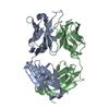
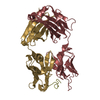
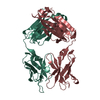
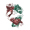
| Details | The asymmetric unit comprises the heavy and light chains that form the Fab fragment |
-Components
| #1: Antibody | Mass: 23868.488 Da / Num. of mol.: 1 Source method: isolated from a genetically manipulated source Source: (gene. exp.)  | ||||||
|---|---|---|---|---|---|---|---|
| #2: Antibody | Mass: 23574.312 Da / Num. of mol.: 1 Source method: isolated from a genetically manipulated source Source: (gene. exp.) | ||||||
| #3: Chemical |   Mass: 92.094 Da / Num. of mol.: 2 / Source method: obtained synthetically / Formula: C3H8O3 Mass: 92.094 Da / Num. of mol.: 2 / Source method: obtained synthetically / Formula: C3H8O3#4: Chemical | ChemComp-MES / |   Mass: 195.237 Da / Num. of mol.: 1 / Source method: obtained synthetically / Formula: C6H13NO4S / Comment: pH buffer*YM Mass: 195.237 Da / Num. of mol.: 1 / Source method: obtained synthetically / Formula: C6H13NO4S / Comment: pH buffer*YM#5: Water | ChemComp-HOH / |  Mass: 18.015 Da / Num. of mol.: 612 / Source method: isolated from a natural source / Formula: H2O Mass: 18.015 Da / Num. of mol.: 612 / Source method: isolated from a natural source / Formula: H2OHas protein modification | Y | |
-Experimental details
-Experiment
| Experiment | Method: X-RAY DIFFRACTION / Number of used crystals: 1 |
|---|
- Sample preparation
| Crystal | Density Matthews: 2.8 Å3/Da / Density % sol: 56.4 % |
|---|---|
| Crystal grow | Temperature: 293 K / Method: vapor diffusion, hanging drop / pH: 6.5 Details: PEG MME 5K, ammonium sulfate, MES, pH 6.5, VAPOR DIFFUSION, HANGING DROP, temperature 293K |
-Data collection
| Diffraction | Mean temperature: 95 K |
|---|---|
| Diffraction source | Source: ROTATING ANODE / Type: RIGAKU RUH2R / Wavelength: 1.5418 Å |
| Detector | Type: RIGAKU RAXIS IV / Detector: IMAGE PLATE / Date: Nov 11, 2003 / Details: Osmic Confocal Blue |
| Radiation | Monochromator: Confocal Optics / Protocol: SINGLE WAVELENGTH / Monochromatic (M) / Laue (L): M / Scattering type: x-ray |
| Radiation wavelength | Wavelength: 1.5418 Å / Relative weight: 1 |
| Reflection | Resolution: 1.83→36.7 Å / Num. obs: 43775 / % possible obs: 92.4 % / Observed criterion σ(F): 0 / Observed criterion σ(I): -3 / Redundancy: 3.7 % / Biso Wilson estimate: 22.2 Å2 / Rsym value: 0.065 / Net I/σ(I): 12.9 |
| Reflection shell | Resolution: 1.83→1.9 Å / Redundancy: 1.4 % / Mean I/σ(I) obs: 2.4 / Rsym value: 0.293 / % possible all: 53 |
- Processing
| Software |
| ||||||||||||||||||||||||||||||||||||
|---|---|---|---|---|---|---|---|---|---|---|---|---|---|---|---|---|---|---|---|---|---|---|---|---|---|---|---|---|---|---|---|---|---|---|---|---|---|
| Refinement | Method to determine structure: MOLECULAR REPLACEMENT Starting model: PDB ENTRY 48G7 Resolution: 1.83→36.6 Å / Rfactor Rfree error: 0.004 / Isotropic thermal model: RESTRAINED / Cross valid method: THROUGHOUT / σ(F): 0 / σ(I): -3 / Stereochemistry target values: Engh & Huber
| ||||||||||||||||||||||||||||||||||||
| Solvent computation | Solvent model: FLAT MODEL / Bsol: 61.3753 Å2 / ksol: 0.385562 e/Å3 | ||||||||||||||||||||||||||||||||||||
| Displacement parameters | Biso mean: 29.2 Å2
| ||||||||||||||||||||||||||||||||||||
| Refine analyze |
| ||||||||||||||||||||||||||||||||||||
| Refinement step | Cycle: LAST / Resolution: 1.83→36.6 Å
| ||||||||||||||||||||||||||||||||||||
| Refine LS restraints |
| ||||||||||||||||||||||||||||||||||||
| LS refinement shell | Resolution: 1.83→1.94 Å / Rfactor Rfree error: 0.017 / Total num. of bins used: 6
| ||||||||||||||||||||||||||||||||||||
| Xplor file |
|
