 Movie
Movie Controller


+ Open data

- Basic information
| Entry | Database: PDB / ID: 1t27 | |||||||||
|---|---|---|---|---|---|---|---|---|---|---|
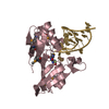
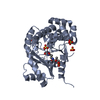
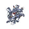
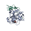
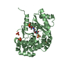
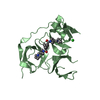
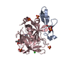
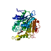
| Title | THE STRUCTURE OF PITP COMPLEXED TO PHOSPHATIDYLCHOLINE | |||||||||
 Components Components | Phosphatidylinositol transfer protein alpha isoform | |||||||||
 Keywords Keywords | LIPID BINDING PROTEIN | |||||||||
| Function / homology |  Function and homology information Function and homology informationstearic acid binding / phosphatidylcholine intramembrane carrier activity / phosphatidylcholine transfer activity / phosphatidylinositol transfer activity / fatty-acyl-CoA binding / phospholipid transport / phosphatidylcholine binding / axonogenesis / phosphatidylinositol binding / phospholipid binding ...stearic acid binding / phosphatidylcholine intramembrane carrier activity / phosphatidylcholine transfer activity / phosphatidylinositol transfer activity / fatty-acyl-CoA binding / phospholipid transport / phosphatidylcholine binding / axonogenesis / phosphatidylinositol binding / phospholipid binding / lipid binding / nucleus / cytoplasm Similarity search - Function | |||||||||
| Biological species |  | |||||||||
| Method |  X-RAY DIFFRACTION / SYNCHROTRON / MAD / Resolution: 2.2 Å X-RAY DIFFRACTION / SYNCHROTRON / MAD / Resolution: 2.2 Å | |||||||||
 Authors Authors | Yoder, M.D. / Thomas, L.M. / Tremblay, J.M. / Oliver, R.L. / Yarbrough, L.R. / Helmkamp Jr., G.M. | |||||||||
 Citation Citation | Journal: J.Biol.Chem. / Year: 2001 Title: STRUCTURE OF A MULTIFUNCTIONAL PROTEIN. MAMMALIAN PHOSPHATIDYLINOSITOL TRANSFER PROTEIN COMPLEXED WITH PHOSPHATIDYLCHOLINE Authors: Yoder, M.D. / Thomas, L.M. / Tremblay, J.M. / Oliver, R.L. / Yarbrough, L.R. / Helmkamp Jr., G.M. #1: Journal: Acta Crystallogr.,Sect.D / Year: 1999 Title: X-RAY ANALYSIS OF CRYSTALS OF RAT PHOSPHATIDYLINOSITOL-TRANSFER PROTEIN WITH BOUND PHOSPHATIDYLCHOLINE Authors: Oliver, R.L. / Tremblay, J.M. / Helmkamp Jr., G.M. / Yarbrough, L.R. / Breakfield, N.W. / Yoder, M.D. | |||||||||
| History |
|
- Structure visualization
| Structure viewer | Molecule: MolmilJmol/JSmol |
|---|
- Downloads & links
-Download
| PDBx/mmCIF format | 1t27.cif.gz | 73.3 KB | Display | PDBx/mmCIF format |
|---|---|---|---|---|
| PDB format | pdb1t27.ent.gz | 53.5 KB | Display | PDB format |
| PDBx/mmJSON format | 1t27.json.gz | Tree view | PDBx/mmJSON format | |
| Others |  Other downloads Other downloads |
-Validation report
| Arichive directory | https://data.pdbj.org/pub/pdb/validation_reports/t2/1t27ftp://data.pdbj.org/pub/pdb/validation_reports/t2/1t27 | HTTPS FTP |
|---|
-Related structure data
| Similar structure data |
|---|


-Links
 PDBj
PDBj- Assembly
| Deposited unit | 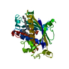
| ||||||||
|---|---|---|---|---|---|---|---|---|---|
| 1 |
| ||||||||
| Unit cell |
|
-Components
| #1: Protein | Mass: 31955.451 Da / Num. of mol.: 1 Source method: isolated from a genetically manipulated source Source: (gene. exp.)  |
|---|---|
| #2: Chemical | ChemComp-PCW /   Mass: 787.121 Da / Num. of mol.: 1 / Source method: obtained synthetically / Formula: C44H85NO8P / Comment: DOPC, phospholipid*YM Mass: 787.121 Da / Num. of mol.: 1 / Source method: obtained synthetically / Formula: C44H85NO8P / Comment: DOPC, phospholipid*YM |
| #3: Water | ChemComp-HOH /  Mass: 18.015 Da / Num. of mol.: 144 / Source method: isolated from a natural source / Formula: H2O Mass: 18.015 Da / Num. of mol.: 144 / Source method: isolated from a natural source / Formula: H2O |
-Experimental details
-Experiment
| Experiment | Method: X-RAY DIFFRACTION / Number of used crystals: 1 |
|---|
- Sample preparation
| Crystal | Density Matthews: 2.25 Å3/Da / Density % sol: 45 % |
|---|---|
| Crystal grow | Temperature: 292 K / Method: vapor diffusion, sitting drop / pH: 8.5 Details: PEG 4000, sodium acetate, Tris-HCL, pH 8.5, VAPOR DIFFUSION, SITTING DROP, temperature 292K |
-Data collection
| Diffraction | Mean temperature: 100 K | ||||||||||||
|---|---|---|---|---|---|---|---|---|---|---|---|---|---|
| Diffraction source | Source: SYNCHROTRON / Site: NSLS  / Beamline: X12C / Wavelength: 0.9791, 0.9794, 0.9500 / Beamline: X12C / Wavelength: 0.9791, 0.9794, 0.9500 | ||||||||||||
| Detector | Type: BRANDEIS - B4 / Detector: CCD / Date: Nov 18, 1999 | ||||||||||||
| Radiation | Monochromator: Si 111 CHANNEL / Protocol: MAD / Monochromatic (M) / Laue (L): M / Scattering type: x-ray | ||||||||||||
| Radiation wavelength |
| ||||||||||||
| Reflection | Resolution: 2.2→50 Å / Num. all: 38629 / Num. obs: 38629 / % possible obs: 90.1 % / Observed criterion σ(F): 0 / Observed criterion σ(I): 0 / Biso Wilson estimate: 7.4 Å2 / Rmerge(I) obs: 0.045 / Net I/σ(I): 20.2 | ||||||||||||
| Reflection shell | Resolution: 2.2→50 Å / Rmerge(I) obs: 0.081 / % possible all: 67.2 |
- Processing
| Software |
| ||||||||||||||||||||||||||||||||||||
|---|---|---|---|---|---|---|---|---|---|---|---|---|---|---|---|---|---|---|---|---|---|---|---|---|---|---|---|---|---|---|---|---|---|---|---|---|---|
| Refinement | Method to determine structure: MAD / Resolution: 2.2→19.94 Å / Rfactor Rfree error: 0.01 / Data cutoff high absF: 1407892.84 / Data cutoff low absF: 0 / Isotropic thermal model: RESTRAINED / Cross valid method: THROUGHOUT / σ(F): 0 / σ(I): 0 / Stereochemistry target values: Engh & Huber
| ||||||||||||||||||||||||||||||||||||
| Solvent computation | Solvent model: FLAT MODEL / Bsol: 36.3224 Å2 / ksol: 0.338776 e/Å3 | ||||||||||||||||||||||||||||||||||||
| Displacement parameters | Biso mean: 21.2 Å2
| ||||||||||||||||||||||||||||||||||||
| Refine analyze |
| ||||||||||||||||||||||||||||||||||||
| Refinement step | Cycle: LAST / Resolution: 2.2→19.94 Å
| ||||||||||||||||||||||||||||||||||||
| Refine LS restraints |
| ||||||||||||||||||||||||||||||||||||
| LS refinement shell | Resolution: 2.2→2.34 Å / Rfactor Rfree error: 0.035 / Total num. of bins used: 6
| ||||||||||||||||||||||||||||||||||||
| Xplor file |
|
