 Movie
Movie Controller

[English] 日本語
 Yorodumi
- PDB-1t0l: Crystal structure of human cytosolic NADP(+)-dependent isocitrate... -
+ Open data

- Basic information
| Entry | Database: PDB / ID: 1t0l | ||||||
|---|---|---|---|---|---|---|---|
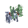
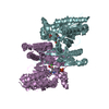
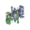
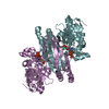
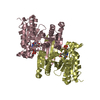
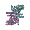
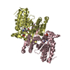
| Title | Crystal structure of human cytosolic NADP(+)-dependent isocitrate dehydrogenase in complex with NADP, isocitrate, and calcium(2+) | ||||||
 Components Components | Isocitrate dehydrogenase [NADP] cytoplasmic | ||||||
 Keywords Keywords | OXIDOREDUCTASE / Rossmann fold / NADP / isocitrate / calcium / quaternary complex | ||||||
| Function / homology |  Function and homology information Function and homology informationAbnormal conversion of 2-oxoglutarate to 2-hydroxyglutarate / NADPH regeneration / regulation of phospholipid catabolic process / regulation of phospholipid biosynthetic process / NFE2L2 regulating TCA cycle genes / isocitrate metabolic process / isocitrate dehydrogenase (NADP+) / NADPH regeneration / isocitrate dehydrogenase (NADP+) activity / NADP+ metabolic process ...Abnormal conversion of 2-oxoglutarate to 2-hydroxyglutarate / NADPH regeneration / regulation of phospholipid catabolic process / regulation of phospholipid biosynthetic process / NFE2L2 regulating TCA cycle genes / isocitrate metabolic process / isocitrate dehydrogenase (NADP+) / NADPH regeneration / isocitrate dehydrogenase (NADP+) activity / NADP+ metabolic process / 2-oxoglutarate metabolic process / glyoxylate cycle / response to steroid hormone / female gonad development / peroxisomal matrix / tricarboxylic acid cycle / glutathione metabolic process / Peroxisomal protein import / NAD binding / NADP binding / tertiary granule lumen / peroxisome / response to oxidative stress / secretory granule lumen / ficolin-1-rich granule lumen / cadherin binding / Neutrophil degranulation / magnesium ion binding / protein homodimerization activity / mitochondrion / extracellular exosome / extracellular region / identical protein binding / cytosol / cytoplasm Similarity search - Function | ||||||
| Biological species |  Homo sapiens (human) Homo sapiens (human) | ||||||
| Method |  X-RAY DIFFRACTION / MOLECULAR REPLACEMENT / Resolution: 2.41 Å X-RAY DIFFRACTION / MOLECULAR REPLACEMENT / Resolution: 2.41 Å | ||||||
 Authors Authors | Xu, X. / Zhao, J. / Peng, B. / Huang, Q. / Arnold, E. / Ding, J. | ||||||
 Citation Citation | Journal: J.Biol.Chem. / Year: 2004 Title: Structures of human cytosolic NADP-dependent isocitrate dehydrogenase reveal a novel self-regulatory mechanism of activity Authors: Xu, X. / Zhao, J. / Xu, Z. / Peng, B. / Huang, Q. / Arnold, E. / Ding, J. | ||||||
| History |
|
- Structure visualization
| Structure viewer | Molecule: MolmilJmol/JSmol |
|---|
- Downloads & links
-Download
| PDBx/mmCIF format | 1t0l.cif.gz | 358.2 KB | Display | PDBx/mmCIF format |
|---|---|---|---|---|
| PDB format | pdb1t0l.ent.gz | 291.5 KB | Display | PDB format |
| PDBx/mmJSON format | 1t0l.json.gz | Tree view | PDBx/mmJSON format | |
| Others |  Other downloads Other downloads |
-Validation report
| Arichive directory | https://data.pdbj.org/pub/pdb/validation_reports/t0/1t0lftp://data.pdbj.org/pub/pdb/validation_reports/t0/1t0l | HTTPS FTP |
|---|
-Related structure data
| Related structure data | 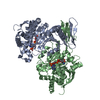 1t09C  1lwdS C: citing same article ( S: Starting model for refinement |
|---|---|
| Similar structure data |


-Links
 PDBj
PDBj


- Assembly
| Deposited unit | 
| ||||||||
|---|---|---|---|---|---|---|---|---|---|
| 1 | 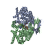
| ||||||||
| 2 | 
| ||||||||
| Unit cell |
|
-Components
| #1: Protein | Mass: 46720.223 Da / Num. of mol.: 4 Source method: isolated from a genetically manipulated source Source: (gene. exp.) Homo sapiens (human) / Gene: IDH / Plasmid: pET-22b(+) / Species (production host): Escherichia coli / Production host:  References: UniProt: O75874, isocitrate dehydrogenase (NADP+) #2: Chemical | ChemComp-CA /   Mass: 40.078 Da / Num. of mol.: 4 / Source method: obtained synthetically / Formula: Ca Mass: 40.078 Da / Num. of mol.: 4 / Source method: obtained synthetically / Formula: Ca#3: Chemical | ChemComp-NAP /   Mass: 743.405 Da / Num. of mol.: 4 / Source method: obtained synthetically / Formula: C21H28N7O17P3 Mass: 743.405 Da / Num. of mol.: 4 / Source method: obtained synthetically / Formula: C21H28N7O17P3#4: Chemical | ChemComp-ICT /   Mass: 192.124 Da / Num. of mol.: 4 / Source method: obtained synthetically / Formula: C6H8O7 Mass: 192.124 Da / Num. of mol.: 4 / Source method: obtained synthetically / Formula: C6H8O7#5: Water | ChemComp-HOH / |  Mass: 18.015 Da / Num. of mol.: 914 / Source method: isolated from a natural source / Formula: H2O Mass: 18.015 Da / Num. of mol.: 914 / Source method: isolated from a natural source / Formula: H2O |
|---|
-Experimental details
-Experiment
| Experiment | Method: X-RAY DIFFRACTION / Number of used crystals: 1 |
|---|
- Sample preparation
| Crystal | Density Matthews: 2.65 Å3/Da / Density % sol: 53.7 % |
|---|---|
| Crystal grow | Temperature: 293 K / Method: vapor diffusion, hanging drop / pH: 5.9 Details: PEG 6000, MES, NADP, isocitrate, calcium chloride, pH 5.9, VAPOR DIFFUSION, HANGING DROP, temperature 293K |
-Data collection
| Diffraction | Mean temperature: 100 K |
|---|---|
| Diffraction source | Source: ROTATING ANODE / Type: RIGAKU ULTRAX 18 / Wavelength: 1.5418 Å |
| Detector | Type: RIGAKU RAXIS IV / Detector: IMAGE PLATE / Date: Dec 30, 2003 / Details: confocal mirror |
| Radiation | Monochromator: confocal mirror / Protocol: SINGLE WAVELENGTH / Monochromatic (M) / Laue (L): M / Scattering type: x-ray |
| Radiation wavelength | Wavelength: 1.5418 Å / Relative weight: 1 |
| Reflection | Resolution: 2.41→12 Å / Num. all: 71234 / Num. obs: 71234 / % possible obs: 95.7 % / Observed criterion σ(F): 0 / Observed criterion σ(I): 0 / Redundancy: 3.1 % / Biso Wilson estimate: 26.1 Å2 / Rmerge(I) obs: 0.085 / Net I/σ(I): 7.8 |
| Reflection shell | Resolution: 2.41→2.5 Å / Redundancy: 2.9 % / Rmerge(I) obs: 0.085 / Mean I/σ(I) obs: 2.9 / Num. unique all: 6671 / % possible all: 95.7 |
- Processing
| Software |
| |||||||||||||||||||||||||||||||||||||||||||||||||||||||||||||||
|---|---|---|---|---|---|---|---|---|---|---|---|---|---|---|---|---|---|---|---|---|---|---|---|---|---|---|---|---|---|---|---|---|---|---|---|---|---|---|---|---|---|---|---|---|---|---|---|---|---|---|---|---|---|---|---|---|---|---|---|---|---|---|---|---|
| Refinement | Method to determine structure: MOLECULAR REPLACEMENT Starting model: PDB ENTRY 1LWD Resolution: 2.41→12 Å / Rfactor Rfree error: 0.004 / Data cutoff high absF: 1576191.17 / Data cutoff low absF: 0 / Isotropic thermal model: RESTRAINED / Cross valid method: THROUGHOUT / σ(F): 0 / Stereochemistry target values: Engh & Huber
| |||||||||||||||||||||||||||||||||||||||||||||||||||||||||||||||
| Solvent computation | Solvent model: FLAT MODEL / Bsol: 76.9338 Å2 / ksol: 0.469785 e/Å3 | |||||||||||||||||||||||||||||||||||||||||||||||||||||||||||||||
| Displacement parameters | Biso mean: 23.7 Å2
| |||||||||||||||||||||||||||||||||||||||||||||||||||||||||||||||
| Refine analyze |
| |||||||||||||||||||||||||||||||||||||||||||||||||||||||||||||||
| Refinement step | Cycle: LAST / Resolution: 2.41→12 Å
| |||||||||||||||||||||||||||||||||||||||||||||||||||||||||||||||
| Refine LS restraints |
| |||||||||||||||||||||||||||||||||||||||||||||||||||||||||||||||
| LS refinement shell | Refine-ID: X-RAY DIFFRACTION / Total num. of bins used: 6
| |||||||||||||||||||||||||||||||||||||||||||||||||||||||||||||||
| Xplor file |
|
