 Movie
Movie Controller

[English] 日本語
 Yorodumi
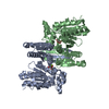
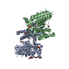
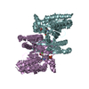
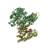
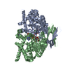
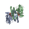
- PDB-1lwd: CRYSTAL STRUCTURE OF NADP-DEPENDENT ISOCITRATE DEHYDROGENASE FROM... -
+ Open data

- Basic information
| Entry | Database: PDB / ID: 1lwd | ||||||
|---|---|---|---|---|---|---|---|
| Title | CRYSTAL STRUCTURE OF NADP-DEPENDENT ISOCITRATE DEHYDROGENASE FROM PORCINE HEART MITOCHONDRIA | ||||||
 Components Components | Isocitrate Dehydrogenase | ||||||
 Keywords Keywords | OXIDOREDUCTASE / TRICARBOXYLIC ACID CYCLE / NADP | ||||||
| Function / homology |  Function and homology information Function and homology informationisocitrate metabolic process / isocitrate dehydrogenase (NADP+) / isocitrate dehydrogenase (NADP+) activity / NADP+ metabolic process / 2-oxoglutarate metabolic process / glyoxylate cycle / tricarboxylic acid cycle / NAD binding / magnesium ion binding / mitochondrion Similarity search - Function | ||||||
| Biological species |  | ||||||
| Method |  X-RAY DIFFRACTION / MAD / Resolution: 1.85 Å X-RAY DIFFRACTION / MAD / Resolution: 1.85 Å | ||||||
 Authors Authors | Ceccarelli, C. / Bahnson, B.J. | ||||||
 Citation Citation | Journal: J.Biol.Chem. / Year: 2002 Title: Crystal Structure of Porcine Mitochondrial NADP+-Dependent Isocitrate Dehydrogenase Complexed with Mn2+ and Isocitrate Authors: Ceccarelli, C. / Grodsky, N.B. / Ariyaratne, N. / Colman, R.F. / Bahnson, B.J. | ||||||
| History |
|
- Structure visualization
| Structure viewer | Molecule: MolmilJmol/JSmol |
|---|
- Downloads & links
-Download
| PDBx/mmCIF format | 1lwd.cif.gz | 191.1 KB | Display | PDBx/mmCIF format |
|---|---|---|---|---|
| PDB format | pdb1lwd.ent.gz | 150.8 KB | Display | PDB format |
| PDBx/mmJSON format | 1lwd.json.gz | Tree view | PDBx/mmJSON format | |
| Others |  Other downloads Other downloads |
-Validation report
| Arichive directory | https://data.pdbj.org/pub/pdb/validation_reports/lw/1lwdftp://data.pdbj.org/pub/pdb/validation_reports/lw/1lwd | HTTPS FTP |
|---|
-Related structure data
| Similar structure data |
|---|


-Links
 PDBj
PDBj


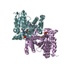
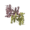
- Assembly
| Deposited unit | 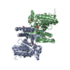
| |||||||||||||||
|---|---|---|---|---|---|---|---|---|---|---|---|---|---|---|---|---|
| 1 |
| |||||||||||||||
| Unit cell |
| |||||||||||||||
| Components on special symmetry positions |
| |||||||||||||||
| Noncrystallographic symmetry (NCS) | NCS oper: (Code: given Matrix: (-0.53688, 0.63102, 0.55998), Vector: Details | THE BIOLOGICALLY FUNCTIONAL MOLECULE IS A DIMER FORMED BY SUBUNITS A AND B, EACH OF WHICH IS PRESENT IN THE CRYSTALLOGRAPHIC ASYMMETRIC UNIT | |
-Components
| #1: Protein | Mass: 46695.219 Da / Num. of mol.: 2 Source method: isolated from a genetically manipulated source Source: (gene. exp.)  References: UniProt: P33198, isocitrate dehydrogenase (NADP+) #2: Chemical |   Mass: 54.938 Da / Num. of mol.: 2 / Source method: obtained synthetically / Formula: Mn Mass: 54.938 Da / Num. of mol.: 2 / Source method: obtained synthetically / Formula: Mn#3: Chemical | 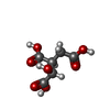  Mass: 192.124 Da / Num. of mol.: 2 / Source method: obtained synthetically / Formula: C6H8O7 Mass: 192.124 Da / Num. of mol.: 2 / Source method: obtained synthetically / Formula: C6H8O7#4: Chemical |   Mass: 96.063 Da / Num. of mol.: 2 / Source method: obtained synthetically / Formula: SO4 Mass: 96.063 Da / Num. of mol.: 2 / Source method: obtained synthetically / Formula: SO4#5: Water | ChemComp-HOH / |  Mass: 18.015 Da / Num. of mol.: 715 / Source method: isolated from a natural source / Formula: H2O Mass: 18.015 Da / Num. of mol.: 715 / Source method: isolated from a natural source / Formula: H2O |
|---|
-Experimental details
-Experiment
| Experiment | Method: X-RAY DIFFRACTION / Number of used crystals: 1 |
|---|
- Sample preparation
| Crystal | Density Matthews: 2.73 Å3/Da / Density % sol: 55 % Description: THE STRUCTURE OF THE SE-MET CRYSTAL WAS SOLVED BY MULTIWAVELENGTH ANOMALOUS DIFFRACTION (MAD). THE THREE-WAVELENGTH MAD EXPERIMENT WAS PERFORMED ON BEAMLINE X12-C AT THE NSLS, BROOKHAVEN NATIONAL LABORATORY. | |||||||||||||||||||||||||||||||||||||||||||||||||||||||||||||||||||||||||||||
|---|---|---|---|---|---|---|---|---|---|---|---|---|---|---|---|---|---|---|---|---|---|---|---|---|---|---|---|---|---|---|---|---|---|---|---|---|---|---|---|---|---|---|---|---|---|---|---|---|---|---|---|---|---|---|---|---|---|---|---|---|---|---|---|---|---|---|---|---|---|---|---|---|---|---|---|---|---|---|
| Crystal grow | Temperature: 277 K / Method: vapor diffusion, hanging drop / pH: 7.7 Details: 20 MG/ML PROTEIN, 100 MM TRIETHANOLAMINE CHLORIDE PH 7.7, 150 MM SODIUM SULFATE, 8 MM ISOCITRATE, 4 MM MANGANESE SULFATE, 20% PEG 6000, 3% GLYCEROL, pH 7.70, VAPOR DIFFUSION, HANGING DROP, temperature 277K | |||||||||||||||||||||||||||||||||||||||||||||||||||||||||||||||||||||||||||||
| Crystal grow | *PLUS Temperature: 4 ℃ / pH: 7.7 | |||||||||||||||||||||||||||||||||||||||||||||||||||||||||||||||||||||||||||||
| Components of the solutions | *PLUS
|
-Data collection
| Diffraction | Mean temperature: 110 K |
|---|---|
| Diffraction source | Source: ROTATING ANODE / Type: RIGAKU RUH3R / Wavelength: 1.5418 / Wavelength: 1.5418 Å |
| Detector | Type: RIGAKU RAXIS IV / Detector: IMAGE PLATE / Date: Dec 1, 1999 / Details: OSMIC MIRRORS |
| Radiation | Protocol: SINGLE WAVELENGTH / Monochromatic (M) / Laue (L): M / Scattering type: x-ray |
| Radiation wavelength | Wavelength: 1.5418 Å / Relative weight: 1 |
| Reflection | Resolution: 1.8→30 Å / Num. all: 88689 / Num. obs: 88689 / % possible obs: 95.9 % / Observed criterion σ(F): 0 / Observed criterion σ(I): 0 / Redundancy: 3.767 % / Biso Wilson estimate: 22.9 Å2 / Rmerge(I) obs: 0.073 / Net I/σ(I): 11.6 |
| Reflection shell | Resolution: 1.8→1.86 Å / Redundancy: 3.643 % / Rmerge(I) obs: 0.963 / Mean I/σ(I) obs: 1.4 / Num. unique all: 8584 / % possible all: 92.7 |
| Reflection | *PLUS Highest resolution: 1.8 Å / Lowest resolution: 30 Å / Num. measured all: 334110 |
- Processing
| Software |
| ||||||||||||||||||||||||||||||||||||
|---|---|---|---|---|---|---|---|---|---|---|---|---|---|---|---|---|---|---|---|---|---|---|---|---|---|---|---|---|---|---|---|---|---|---|---|---|---|
| Refinement | Method to determine structure: MAD Starting model: ISOMORPHOUS SE-MET CRYSTAL STRUCTURE Resolution: 1.85→29.23 Å / Rfactor Rfree error: 0.002 / Isotropic thermal model: RESTRAINED / Cross valid method: THROUGHOUT / σ(F): 0 / Stereochemistry target values: Engh & Huber Details: NCS RESTRAINTS WERE APPLIED TO 3074 OF 3285 PAIRS OF ATOMS IN THE A AND B SUBUNITS OF THE PROTEIN. SEE REMARK 295 FOR RESIDUES AND SIDE CHAIN ATOMS OMITTED FROM NCS RESTRAINTS.
| ||||||||||||||||||||||||||||||||||||
| Solvent computation | Solvent model: FLAT MODEL / Bsol: 46.6925 Å2 / ksol: 0.352708 e/Å3 | ||||||||||||||||||||||||||||||||||||
| Displacement parameters | Biso mean: 27.4 Å2
| ||||||||||||||||||||||||||||||||||||
| Refine analyze |
| ||||||||||||||||||||||||||||||||||||
| Refinement step | Cycle: LAST / Resolution: 1.85→29.23 Å
| ||||||||||||||||||||||||||||||||||||
| Refine LS restraints |
| ||||||||||||||||||||||||||||||||||||
| Refine LS restraints NCS | NCS model details: RESTRAINED / Rms dev Biso : 1.936 Å2 / Rms dev position: 0.066 Å / Weight Biso : 2 / Weight position: 50 | ||||||||||||||||||||||||||||||||||||
| LS refinement shell | Resolution: 1.85→1.97 Å / Rfactor Rfree error: 0.009 / Total num. of bins used: 6
| ||||||||||||||||||||||||||||||||||||
| Xplor file |
| ||||||||||||||||||||||||||||||||||||
| Refinement | *PLUS % reflection Rfree: 10 % / Rfactor Rfree: 0.21 | ||||||||||||||||||||||||||||||||||||
| Solvent computation | *PLUS | ||||||||||||||||||||||||||||||||||||
| Displacement parameters | *PLUS | ||||||||||||||||||||||||||||||||||||
| Refine LS restraints | *PLUS
|
