 Movie
Movie Controller


+ Open data

- Basic information
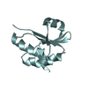
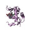
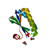
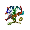
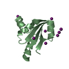
| Entry | Database: PDB / ID: 1svy | ||||||
|---|---|---|---|---|---|---|---|
| Title | SEVERIN DOMAIN 2, 1.75 ANGSTROM CRYSTAL STRUCTURE | ||||||
 Components Components | SEVERIN | ||||||
 Keywords Keywords | ACTIN-BINDING PROTEIN / CALCIUM-BINDING / CYTOSKELETON / GELSOLIN / SEVERIN / VILLIN / CALCIUM / PIP2 | ||||||
| Function / homology |  Function and homology information Function and homology informationactin filament severing activity / actin filament fragmentation / actin filament severing / barbed-end actin filament capping / actin polymerization or depolymerization / actin filament bundle assembly / actin monomer binding / phagocytic vesicle / phosphatidylinositol-4,5-bisphosphate binding / calcium-dependent protein binding ...actin filament severing activity / actin filament fragmentation / actin filament severing / barbed-end actin filament capping / actin polymerization or depolymerization / actin filament bundle assembly / actin monomer binding / phagocytic vesicle / phosphatidylinositol-4,5-bisphosphate binding / calcium-dependent protein binding / actin filament binding / actin cytoskeleton / calcium ion binding Similarity search - Function | ||||||
| Biological species |  | ||||||
| Method |  X-RAY DIFFRACTION / SYNCHROTRON / MIRAS / Resolution: 1.75 Å X-RAY DIFFRACTION / SYNCHROTRON / MIRAS / Resolution: 1.75 Å | ||||||
 Authors Authors | Puius, Y.A. / Fedorov, E.V. / Eichinger, L. / Sullivan, M. / Schleicher, M. / Almo, S.C. | ||||||
 Citation Citation | Journal: Biochemistry / Year: 2000 Title: Mapping the functional surface of domain 2 in the gelsolin superfamily. Authors: Puius, Y.A. / Fedorov, E.V. / Eichinger, L. / Schleicher, M. / Almo, S.C. #1: Journal: J.Mol.Biol. / Year: 1995Title: Structure of Severin Domain 2 in Solution Authors: Schnuchel, A. / Wiltscheck, R. / Eichinger, L. / Schleicher, M. / Holak, T.A. #2: Journal: Biochemistry / Year: 1992Title: Characterization of Actin-and Lipid-Binding Domains in Severin, a Ca(2+)-Dependent F-Actin Fragmenting Protein Authors: Eichinger, L. / Schleicher, M. #3: Journal: J.Biol.Chem. / Year: 1988Title: Severin, Gelsolin, and Villin Share a Homologous Sequence in Regions Presumed to Contain F-Actin Severing Domains Authors: Andre, E. / Lottspeich, F. / Schleicher, M. / Noegel, A. | ||||||
| History |
|
- Structure visualization
| Structure viewer | Molecule: MolmilJmol/JSmol |
|---|
- Downloads & links
-Download
| PDBx/mmCIF format | 1svy.cif.gz | 36.2 KB | Display | PDBx/mmCIF format |
|---|---|---|---|---|
| PDB format | pdb1svy.ent.gz | 23.1 KB | Display | PDB format |
| PDBx/mmJSON format | 1svy.json.gz | Tree view | PDBx/mmJSON format | |
| Others |  Other downloads Other downloads |
-Validation report
| Arichive directory | https://data.pdbj.org/pub/pdb/validation_reports/sv/1svyftp://data.pdbj.org/pub/pdb/validation_reports/sv/1svy | HTTPS FTP |
|---|
-Related structure data
| Similar structure data |
|---|


-Links
 PDBj
PDBj
- Assembly
| Deposited unit | 
| ||||||||
|---|---|---|---|---|---|---|---|---|---|
| 1 |
| ||||||||
| Unit cell |
|
-Components
| #1: Protein | Mass: 12276.842 Da / Num. of mol.: 1 / Fragment: DOMAIN 2 Source method: isolated from a genetically manipulated source Source: (gene. exp.)  |
|---|---|
| #2: Chemical | ChemComp-CA /   Mass: 40.078 Da / Num. of mol.: 1 / Source method: obtained synthetically / Formula: Ca Mass: 40.078 Da / Num. of mol.: 1 / Source method: obtained synthetically / Formula: Ca |
| #3: Chemical | ChemComp-NA /   Mass: 22.990 Da / Num. of mol.: 1 / Source method: obtained synthetically / Formula: Na Mass: 22.990 Da / Num. of mol.: 1 / Source method: obtained synthetically / Formula: Na |
| #4: Water | ChemComp-HOH /  Mass: 18.015 Da / Num. of mol.: 114 / Source method: isolated from a natural source / Formula: H2O Mass: 18.015 Da / Num. of mol.: 114 / Source method: isolated from a natural source / Formula: H2O |
| Has protein modification | Y |
-Experimental details
-Experiment
| Experiment | Method: X-RAY DIFFRACTION / Number of used crystals: 1 |
|---|
- Sample preparation
| Crystal | Density Matthews: 2.11 Å3/Da / Density % sol: 42 % | ||||||||||||||||||||
|---|---|---|---|---|---|---|---|---|---|---|---|---|---|---|---|---|---|---|---|---|---|
| Crystal grow | pH: 8 / Details: pH 8.0 | ||||||||||||||||||||
| Crystal | *PLUS | ||||||||||||||||||||
| Crystal grow | *PLUS Method: vapor diffusion, hanging drop | ||||||||||||||||||||
| Components of the solutions | *PLUS
|
-Data collection
| Diffraction | Mean temperature: 140 K |
|---|---|
| Diffraction source | Source: SYNCHROTRON / Site: NSLS  / Beamline: X9B / Wavelength: 1.2 / Beamline: X9B / Wavelength: 1.2 |
| Detector | Type: FUJI / Detector: IMAGE PLATE / Date: Feb 1, 1996 |
| Radiation | Monochromatic (M) / Laue (L): M / Scattering type: x-ray |
| Radiation wavelength | Wavelength: 1.2 Å / Relative weight: 1 |
| Reflection | Resolution: 1.75→20 Å / Num. obs: 10235 / % possible obs: 93.1 % / Observed criterion σ(I): 0 / Rmerge(I) obs: 0.035 / Net I/σ(I): 30.9 |
| Reflection shell | Resolution: 1.75→1.81 Å / Rmerge(I) obs: 0.116 / Mean I/σ(I) obs: 9.9 / % possible all: 77.1 |
| Reflection shell | *PLUS % possible obs: 77.1 % |
- Processing
| Software |
| ||||||||||||||||||||||||||||||||||||||||||||||||||||||||||||
|---|---|---|---|---|---|---|---|---|---|---|---|---|---|---|---|---|---|---|---|---|---|---|---|---|---|---|---|---|---|---|---|---|---|---|---|---|---|---|---|---|---|---|---|---|---|---|---|---|---|---|---|---|---|---|---|---|---|---|---|---|---|
| Refinement | Method to determine structure: MIRAS / Resolution: 1.75→20 Å / Cross valid method: THROUGHOUT / σ(F): 0 / Details: ISOTROPIC SOLVENT MASK
| ||||||||||||||||||||||||||||||||||||||||||||||||||||||||||||
| Displacement parameters | Biso mean: 20.1 Å2 | ||||||||||||||||||||||||||||||||||||||||||||||||||||||||||||
| Refinement step | Cycle: LAST / Resolution: 1.75→20 Å
| ||||||||||||||||||||||||||||||||||||||||||||||||||||||||||||
| Refine LS restraints |
| ||||||||||||||||||||||||||||||||||||||||||||||||||||||||||||
| LS refinement shell | Resolution: 1.75→1.81 Å / Total num. of bins used: 10
| ||||||||||||||||||||||||||||||||||||||||||||||||||||||||||||
| Software | *PLUS Name: X-PLOR / Version: 3.851 / Classification: refinement | ||||||||||||||||||||||||||||||||||||||||||||||||||||||||||||
| Refinement | *PLUS Rfactor Rfree: 0.2462 | ||||||||||||||||||||||||||||||||||||||||||||||||||||||||||||
| Solvent computation | *PLUS | ||||||||||||||||||||||||||||||||||||||||||||||||||||||||||||
| Displacement parameters | *PLUS | ||||||||||||||||||||||||||||||||||||||||||||||||||||||||||||
| LS refinement shell | *PLUS Rfactor obs: 0.274 |
