 Movie
Movie Controller

[English] 日本語
 Yorodumi
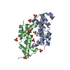
- PDB-1sqe: 1.5A Crystal Structure Of the protein PG130 from Staphylococcus a... -
+ Open data

- Basic information
| Entry | Database: PDB / ID: 1sqe | ||||||
|---|---|---|---|---|---|---|---|
| Title | 1.5A Crystal Structure Of the protein PG130 from Staphylococcus aureus, Structural genomics | ||||||
 Components Components | hypothetical protein PG130 | ||||||
 Keywords Keywords | STRUCTURAL GENOMICS / UNKNOWN FUNCTION / PSI / Protein Structure Initiative / Midwest Center for Structural Genomics / MCSG | ||||||
| Function / homology |  Function and homology information Function and homology informationheme oxygenase (staphylobilin-producing) / iron import into cell / heme oxygenase (decyclizing) activity / heme catabolic process / iron ion binding / heme binding / cytoplasm Similarity search - Function | ||||||
| Biological species |   Staphylococcus aureus (bacteria) Staphylococcus aureus (bacteria) | ||||||
| Method |  X-RAY DIFFRACTION / SYNCHROTRON / MAD / Resolution: 1.5 Å X-RAY DIFFRACTION / SYNCHROTRON / MAD / Resolution: 1.5 Å | ||||||
 Authors Authors | Zhang, R. / Wu, R. / Joachimiak, G. / Schneewind, O. / Joachimiak, A. / Midwest Center for Structural Genomics (MCSG) | ||||||
 Citation Citation | Journal: J.Biol.Chem. / Year: 2005 Title: Staphylococcus aureus IsdG and IsdI, heme-degrading enzymes with structural similarity to monooxygenases Authors: Wu, R. / Skaar, E.P. / Zhang, R. / Joachimiak, G. / Gornicki, P. / Schneewind, O. / Joachimiak, A. | ||||||
| History |
|
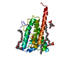
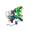
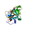
- Structure visualization
| Structure viewer | Molecule: MolmilJmol/JSmol |
|---|
- Downloads & links
-Download
| PDBx/mmCIF format | 1sqe.cif.gz | 59.1 KB | Display | PDBx/mmCIF format |
|---|---|---|---|---|
| PDB format | pdb1sqe.ent.gz | 43.4 KB | Display | PDB format |
| PDBx/mmJSON format | 1sqe.json.gz | Tree view | PDBx/mmJSON format | |
| Others |  Other downloads Other downloads |
-Validation report
| Arichive directory | https://data.pdbj.org/pub/pdb/validation_reports/sq/1sqeftp://data.pdbj.org/pub/pdb/validation_reports/sq/1sqe | HTTPS FTP |
|---|
-Related structure data


-Links
 PDBj
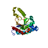
PDBj- Assembly
| Deposited unit | 
| ||||||||
|---|---|---|---|---|---|---|---|---|---|
| 1 |
| ||||||||
| Unit cell |
| ||||||||
| Details | This protein existed as dimer. MolA and MolB represents the dimer in asymmetric unit. |
-Components
| #1: Protein | Mass: 12944.316 Da / Num. of mol.: 2 Source method: isolated from a genetically manipulated source Source: (gene. exp.) Staphylococcus aureus (bacteria) / Strain: MU50 / Plasmid: PDM68 / Species (production host): Escherichia coli / Production host: #2: Water | ChemComp-HOH / |  Mass: 18.015 Da / Num. of mol.: 231 / Source method: isolated from a natural source / Formula: H2O Mass: 18.015 Da / Num. of mol.: 231 / Source method: isolated from a natural source / Formula: H2O |
|---|
-Experimental details
-Experiment
| Experiment | Method: X-RAY DIFFRACTION / Number of used crystals: 1 |
|---|
- Sample preparation
| Crystal | Density Matthews: 2.04 Å3/Da / Density % sol: 39.68 % Description: FRIEDEL PAIRS WERE USED IN DETERMINING THIS STRUCTURE |
|---|---|
| Crystal grow | Temperature: 289 K / Method: vapor diffusion, hanging drop / pH: 6.5 Details: 25% PEG 4000, 0.25M NaCl, pH 6.5, VAPOR DIFFUSION, HANGING DROP, temperature 289K |
-Data collection
| Diffraction | Mean temperature: 100 K | ||||||||||||
|---|---|---|---|---|---|---|---|---|---|---|---|---|---|
| Diffraction source | Source: SYNCHROTRON / Site: APS  / Beamline: 19-ID / Wavelength: 0.9795, 0.9798, 0.94656 / Beamline: 19-ID / Wavelength: 0.9795, 0.9798, 0.94656 | ||||||||||||
| Detector | Type: SBC-2 / Detector: CCD / Date: Oct 26, 2003 / Details: mirrors | ||||||||||||
| Radiation | Monochromator: Si(111) / Protocol: MAD / Monochromatic (M) / Laue (L): M / Scattering type: x-ray | ||||||||||||
| Radiation wavelength |
| ||||||||||||
| Reflection | Resolution: 1.5→50 Å / Num. all: 65616 / Num. obs: 59383 / % possible obs: 94.7 % / Observed criterion σ(F): 4 / Observed criterion σ(I): 4 / Redundancy: 4.78 % / Biso Wilson estimate: 15.7 Å2 / Rmerge(I) obs: 0.087 / Net I/σ(I): 23.29 | ||||||||||||
| Reflection shell | Resolution: 1.5→1.55 Å / Redundancy: 3.8 % / Rmerge(I) obs: 0.274 / Mean I/σ(I) obs: 2.12 / Num. unique all: 2589 / % possible all: 77.4 |
- Processing
| Software |
| |||||||||||||||||||||||||
|---|---|---|---|---|---|---|---|---|---|---|---|---|---|---|---|---|---|---|---|---|---|---|---|---|---|---|
| Refinement | Method to determine structure: MAD / Resolution: 1.5→19.4 Å / Rfactor Rfree error: 0.005 / Data cutoff high absF: 360920.52 / Data cutoff low absF: 0 / Isotropic thermal model: RESTRAINED / Cross valid method: THROUGHOUT / σ(F): 0 / Stereochemistry target values: Engh & Huber Details: FRIEDEL PAIRS WERE USED IN DETERMINING THIS STRUCTURE
| |||||||||||||||||||||||||
| Solvent computation | Solvent model: FLAT MODEL / Bsol: 46.2343 Å2 / ksol: 0.362331 e/Å3 | |||||||||||||||||||||||||
| Displacement parameters | Biso mean: 21.5 Å2
| |||||||||||||||||||||||||
| Refine analyze |
| |||||||||||||||||||||||||
| Refinement step | Cycle: LAST / Resolution: 1.5→19.4 Å
| |||||||||||||||||||||||||
| Refine LS restraints |
| |||||||||||||||||||||||||
| LS refinement shell | Resolution: 1.5→1.59 Å / Rfactor Rfree error: 0.015 / Total num. of bins used: 6
| |||||||||||||||||||||||||
| Xplor file |
|
