 Movie
Movie Controller


+ Open data

- Basic information
| Entry | Database: PDB / ID: 3wyi | ||||||
|---|---|---|---|---|---|---|---|
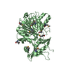
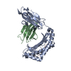
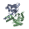
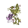
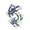
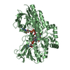
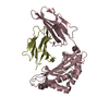
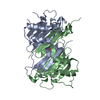
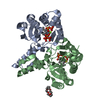
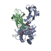
| Title | Structure of S. aureus undecaprenyl diphosphate synthase | ||||||
 Components Components | Isoprenyl transferase | ||||||
 Keywords Keywords | TRANSFERASE / isopentenyl diphosphate binding / product inhibition | ||||||
| Function / homology | Undecaprenyl pyrophosphate synthetase / Decaprenyl diphosphate synthase-like / 3-Layer(aba) Sandwich / Alpha Beta / :  Function and homology information Function and homology information | ||||||
| Biological species |   Staphylococcus aureus (bacteria) Staphylococcus aureus (bacteria) | ||||||
| Method |  X-RAY DIFFRACTION / SYNCHROTRON / MOLECULAR REPLACEMENT / Resolution: 2 Å X-RAY DIFFRACTION / SYNCHROTRON / MOLECULAR REPLACEMENT / Resolution: 2 Å | ||||||
 Authors Authors | Gao, J. / Ko, T.P. / Huang, C.H. / Oldfield, E. / Guo, R.T. | ||||||
 Citation Citation | Journal: J.Med.Chem. / Year: 2015 Title: Antibacterial drug leads: DNA and enzyme multitargeting. Authors: Zhu, W. / Wang, Y. / Li, K. / Gao, J. / Huang, C.H. / Chen, C.C. / Ko, T.P. / Zhang, Y. / Guo, R.T. / Oldfield, E. | ||||||
| History |
|
- Structure visualization
| Structure viewer | Molecule: MolmilJmol/JSmol |
|---|
- Downloads & links
-Download
| PDBx/mmCIF format | 3wyi.cif.gz | 69.8 KB | Display | PDBx/mmCIF format |
|---|---|---|---|---|
| PDB format | pdb3wyi.ent.gz | 50.4 KB | Display | PDB format |
| PDBx/mmJSON format | 3wyi.json.gz | Tree view | PDBx/mmJSON format | |
| Others |  Other downloads Other downloads |
-Validation report
| Arichive directory | https://data.pdbj.org/pub/pdb/validation_reports/wy/3wyiftp://data.pdbj.org/pub/pdb/validation_reports/wy/3wyi | HTTPS FTP |
|---|
-Related structure data
| Related structure data |  3wyjC  4u82C  4u8aC  4u8bC  4u8cC  4h8eS C: citing same article ( S: Starting model for refinement |
|---|---|
| Similar structure data |
-Links
 PDBj
PDBj- Assembly
| Deposited unit | 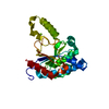
| ||||||||
|---|---|---|---|---|---|---|---|---|---|
| 1 | 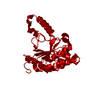
| ||||||||
| Unit cell |
|
-Components
| #1: Protein | Mass: 29902.199 Da / Num. of mol.: 1 Source method: isolated from a genetically manipulated source Source: (gene. exp.) Staphylococcus aureus (bacteria) / Gene: CH52_12785 / Plasmid: pET46Ek/LIC / Production host: References: UniProt: W8UYA6, Transferases; Transferring alkyl or aryl groups, other than methyl groups | ||
|---|---|---|---|
| #2: Chemical |   Mass: 24.305 Da / Num. of mol.: 2 / Source method: obtained synthetically / Formula: Mg Mass: 24.305 Da / Num. of mol.: 2 / Source method: obtained synthetically / Formula: Mg#3: Water | ChemComp-HOH / |  Mass: 18.015 Da / Num. of mol.: 375 / Source method: isolated from a natural source / Formula: H2O Mass: 18.015 Da / Num. of mol.: 375 / Source method: isolated from a natural source / Formula: H2O |
-Experimental details
-Experiment
| Experiment | Method: X-RAY DIFFRACTION / Number of used crystals: 1 |
|---|
- Sample preparation
| Crystal | Density Matthews: 2.48 Å3/Da / Density % sol: 50.47 % |
|---|---|
| Crystal grow | Temperature: 295 K / Method: vapor diffusion, sitting drop / pH: 6 Details: 100mM HEPES, pH 6.5, 200mM Magnesium chloride, 200mM Nickel chloride, 20% PEG 3350, VAPOR DIFFUSION, SITTING DROP, temperature 295K |
-Data collection
| Diffraction | Mean temperature: 100 K |
|---|---|
| Diffraction source | Source: SYNCHROTRON / Site: NSRRC  / Beamline: BL13B1 / Wavelength: 1 Å / Beamline: BL13B1 / Wavelength: 1 Å |
| Detector | Type: ADSC QUANTUM 315 / Detector: CCD / Date: May 1, 2013 |
| Radiation | Protocol: SINGLE WAVELENGTH / Monochromatic (M) / Laue (L): M / Scattering type: x-ray |
| Radiation wavelength | Wavelength: 1 Å / Relative weight: 1 |
| Reflection | Resolution: 2→25 Å / Num. obs: 20923 / % possible obs: 100 % / Redundancy: 8.5 % / Rmerge(I) obs: 0.066 / Net I/σ(I): 32.8 |
| Reflection shell | Resolution: 2→2.07 Å / Redundancy: 8.2 % / Rmerge(I) obs: 0.5 / Mean I/σ(I) obs: 3.6 / Num. unique all: 17400 / % possible all: 100 |
- Processing
| Software |
| ||||||||||||||||||||
|---|---|---|---|---|---|---|---|---|---|---|---|---|---|---|---|---|---|---|---|---|---|
| Refinement | Method to determine structure: MOLECULAR REPLACEMENT Starting model: 4H8E Resolution: 2→25 Å / Stereochemistry target values: Engh & Huber
| ||||||||||||||||||||
| Refinement step | Cycle: LAST / Resolution: 2→25 Å
| ||||||||||||||||||||
| LS refinement shell | Resolution: 2→2.07 Å
|
