 Movie
Movie Controller

[English] 日本語
 Yorodumi
- PDB-4h8e: Structure of S. aureus undecaprenyl diphosphate synthase in compl... -
+ Open data

- Basic information
| Entry | Database: PDB / ID: 4h8e | ||||||
|---|---|---|---|---|---|---|---|
| Title | Structure of S. aureus undecaprenyl diphosphate synthase in complex with FPP and sulfate | ||||||
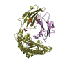
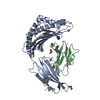
 Components Components | Undecaprenyl pyrophosphate synthase | ||||||
 Keywords Keywords | TRANSFERASE/TRANSFERASE INHIBITOR / alpha-helix / prenyl transferase / cell wall biosynthesis / farnesyl diphosphate binding / isopentenyl diphosphate binding / TRANSFERASE-TRANSFERASE INHIBITOR complex | ||||||
| Function / homology |  Function and homology information Function and homology informationditrans,polycis-undecaprenyl-diphosphate synthase [(2E,6E)-farnesyl-diphosphate specific] activity / polyprenol biosynthetic process / Transferases; Transferring alkyl or aryl groups, other than methyl groups / manganese ion binding / magnesium ion binding / cytosol Similarity search - Function | ||||||
| Biological species |   Staphylococcus aureus subsp. aureus (bacteria) Staphylococcus aureus subsp. aureus (bacteria) | ||||||
| Method |  X-RAY DIFFRACTION / SYNCHROTRON / MOLECULAR REPLACEMENT / Resolution: 1.3 Å X-RAY DIFFRACTION / SYNCHROTRON / MOLECULAR REPLACEMENT / Resolution: 1.3 Å | ||||||
 Authors Authors | Zhu, W. / Oldfield, E. | ||||||
 Citation Citation | Journal: Proc.Natl.Acad.Sci.USA / Year: 2013 Title: Antibacterial drug leads targeting isoprenoid biosynthesis. Authors: Zhu, W. / Zhang, Y. / Sinko, W. / Hensler, M.E. / Olson, J. / Molohon, K.J. / Lindert, S. / Cao, R. / Li, K. / Wang, K. / Wang, Y. / Liu, Y.L. / Sankovsky, A. / de Oliveira, C.A. / Mitchell, ...Authors: Zhu, W. / Zhang, Y. / Sinko, W. / Hensler, M.E. / Olson, J. / Molohon, K.J. / Lindert, S. / Cao, R. / Li, K. / Wang, K. / Wang, Y. / Liu, Y.L. / Sankovsky, A. / de Oliveira, C.A. / Mitchell, D.A. / Nizet, V. / McCammon, J.A. / Oldfield, E. | ||||||
| History |
|
- Structure visualization
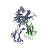
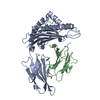
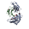
| Structure viewer | Molecule: MolmilJmol/JSmol |
|---|
- Downloads & links
-Download
| PDBx/mmCIF format | 4h8e.cif.gz | 65.3 KB | Display | PDBx/mmCIF format |
|---|---|---|---|---|
| PDB format | pdb4h8e.ent.gz | 47 KB | Display | PDB format |
| PDBx/mmJSON format | 4h8e.json.gz | Tree view | PDBx/mmJSON format | |
| Others |  Other downloads Other downloads |
-Validation report
| Arichive directory | https://data.pdbj.org/pub/pdb/validation_reports/h8/4h8eftp://data.pdbj.org/pub/pdb/validation_reports/h8/4h8e | HTTPS FTP |
|---|
-Related structure data
| Related structure data | 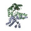 3sgtC  3sgvC  3sgxC  3sh0C  4h2jC  4h2mC  4h2oC  4h38C  4h3aC  4h3cC C: citing same article ( |
|---|---|
| Similar structure data |


-Links
 PDBj
PDBj- Assembly
| Deposited unit | 
| ||||||||
|---|---|---|---|---|---|---|---|---|---|
| 1 | 
| ||||||||
| Unit cell |
|
-Components
| #1: Protein | Mass: 29902.199 Da / Num. of mol.: 1 Source method: isolated from a genetically manipulated source Source: (gene. exp.) Staphylococcus aureus subsp. aureus (bacteria)Strain: N315 / Gene: uppS, SA1103 / Production host: References: UniProt: P60477, ditrans,polycis-undecaprenyl-diphosphate synthase [(2E,6E)-farnesyl-diphosphate specific] |
|---|---|
| #2: Chemical | ChemComp-MG /   Mass: 24.305 Da / Num. of mol.: 1 / Source method: obtained synthetically / Formula: Mg Mass: 24.305 Da / Num. of mol.: 1 / Source method: obtained synthetically / Formula: Mg |
| #3: Chemical | ChemComp-FPP / 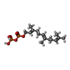  Mass: 382.326 Da / Num. of mol.: 1 / Source method: obtained synthetically / Formula: C15H28O7P2 Mass: 382.326 Da / Num. of mol.: 1 / Source method: obtained synthetically / Formula: C15H28O7P2 |
| #4: Chemical | ChemComp-SO4 /   Mass: 96.063 Da / Num. of mol.: 1 / Source method: obtained synthetically / Formula: SO4 Mass: 96.063 Da / Num. of mol.: 1 / Source method: obtained synthetically / Formula: SO4 |
| #5: Water | ChemComp-HOH /  Mass: 18.015 Da / Num. of mol.: 134 / Source method: isolated from a natural source / Formula: H2O Mass: 18.015 Da / Num. of mol.: 134 / Source method: isolated from a natural source / Formula: H2O |
-Experimental details
-Experiment
| Experiment | Method: X-RAY DIFFRACTION / Number of used crystals: 1 |
|---|
- Sample preparation
| Crystal | Density Matthews: 2.18 Å3/Da / Density % sol: 43.58 % |
|---|---|
| Crystal grow | Temperature: 298 K / Method: vapor diffusion, hanging drop / pH: 7.5 Details: 100 mM MES sodium, pH 6.5, 200 mM ammonium sulfate, 25% PEG5000 MME, VAPOR DIFFUSION, HANGING DROP, temperature 298K |
-Data collection
| Diffraction | Mean temperature: 100 K |
|---|---|
| Diffraction source | Source: SYNCHROTRON / Site: APS  / Beamline: 21-ID-G / Wavelength: 0.97857 Å / Beamline: 21-ID-G / Wavelength: 0.97857 Å |
| Detector | Type: MARMOSAIC 300 mm CCD / Detector: CCD / Date: Jun 16, 2012 |
| Radiation | Monochromator: diamond(111) / Protocol: SINGLE WAVELENGTH / Monochromatic (M) / Laue (L): M / Scattering type: x-ray |
| Radiation wavelength | Wavelength: 0.97857 Å / Relative weight: 1 |
| Reflection | Resolution: 1.3→50 Å / Num. obs: 66126 / % possible obs: 100 % / Observed criterion σ(F): 0 / Observed criterion σ(I): 2 / Redundancy: 13.9 % / Rmerge(I) obs: 0.077 / Net I/σ(I): 33.9 |
| Reflection shell | Resolution: 1.3→1.32 Å / Redundancy: 11.3 % / Rmerge(I) obs: 0.4 / Mean I/σ(I) obs: 5.3 / % possible all: 100 |
- Processing
| Software |
| |||||||||||||||||||||||||||||||||||||||||||||
|---|---|---|---|---|---|---|---|---|---|---|---|---|---|---|---|---|---|---|---|---|---|---|---|---|---|---|---|---|---|---|---|---|---|---|---|---|---|---|---|---|---|---|---|---|---|---|
| Refinement | Method to determine structure: MOLECULAR REPLACEMENT / Resolution: 1.3→32.64 Å / Cor.coef. Fo:Fc: 0.963 / Cor.coef. Fo:Fc free: 0.953 / Occupancy max: 1 / Occupancy min: 0.5 / SU B: 0.565 / SU ML: 0.025 / Cross valid method: THROUGHOUT / σ(F): 0 / ESU R: 0.045 / ESU R Free: 0.047 / Stereochemistry target values: MAXIMUM LIKELIHOOD Details: HYDROGENS HAVE BEEN USED IF PRESENT IN THE INPUT U VALUES : REFINED INDIVIDUALLY
| |||||||||||||||||||||||||||||||||||||||||||||
| Solvent computation | Ion probe radii: 0.8 Å / Shrinkage radii: 0.8 Å / VDW probe radii: 1.2 Å / Solvent model: MASK | |||||||||||||||||||||||||||||||||||||||||||||
| Displacement parameters | Biso max: 56.36 Å2 / Biso mean: 13.6007 Å2 / Biso min: 6.1 Å2
| |||||||||||||||||||||||||||||||||||||||||||||
| Refinement step | Cycle: LAST / Resolution: 1.3→32.64 Å
| |||||||||||||||||||||||||||||||||||||||||||||
| Refine LS restraints |
| |||||||||||||||||||||||||||||||||||||||||||||
| LS refinement shell | Resolution: 1.3→1.334 Å / Total num. of bins used: 20
|
