 Movie
Movie Controller


+ Open data

- Basic information
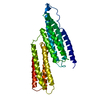
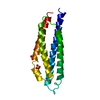
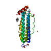
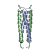
| Entry | Database: PDB / ID: 1sj7 | ||||||
|---|---|---|---|---|---|---|---|
| Title | Crystal Structure of Talin Rod 482-655 | ||||||
 Components Components | Talin 1 | ||||||
 Keywords Keywords | STRUCTURAL PROTEIN | ||||||
| Function / homology |  Function and homology information Function and homology informationGRB2:SOS provides linkage to MAPK signaling for Integrins / Integrin signaling / p130Cas linkage to MAPK signaling for integrins / SEMA3A-Plexin repulsion signaling by inhibiting Integrin adhesion / MAP2K and MAPK activation / LIM domain binding / Smooth Muscle Contraction / Platelet degranulation / cortical microtubule organization / vinculin binding ...GRB2:SOS provides linkage to MAPK signaling for Integrins / Integrin signaling / p130Cas linkage to MAPK signaling for integrins / SEMA3A-Plexin repulsion signaling by inhibiting Integrin adhesion / MAP2K and MAPK activation / LIM domain binding / Smooth Muscle Contraction / Platelet degranulation / cortical microtubule organization / vinculin binding / integrin activation / cell-substrate junction assembly / cortical actin cytoskeleton organization / phosphatidylserine binding / ruffle / phosphatidylinositol binding / integrin-mediated signaling pathway / adherens junction / structural constituent of cytoskeleton / ruffle membrane / integrin binding / actin filament binding / cytoskeleton / cell adhesion / focal adhesion / cell surface / cytosol Similarity search - Function | ||||||
| Biological species |  | ||||||
| Method |  X-RAY DIFFRACTION / SYNCHROTRON / SAD / Resolution: 2.5 Å X-RAY DIFFRACTION / SYNCHROTRON / SAD / Resolution: 2.5 Å | ||||||
 Authors Authors | Papagrigoriou, E. / Gingras, A.R. / Barsukov, I.L. / Critchley, D.R. / Emsley, J. | ||||||
 Citation Citation | Journal: EMBO J. / Year: 2004 Title: Activation of a vinculin-binding site in the talin rod involves rearrangement of a five-helix bundle Authors: Papagrigoriou, E. / Gingras, A.R. / Barsukov, I.L. / Bate, N. / Fillingham, I.J. / Patel, B. / Frank, R. / Ziegler, W.H. / Roberts, G.C. / Critchley, D.R. / Emsley, J. | ||||||
| History |
|
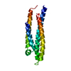
- Structure visualization
| Structure viewer | Molecule: MolmilJmol/JSmol |
|---|
- Downloads & links
-Download
| PDBx/mmCIF format | 1sj7.cif.gz | 102.6 KB | Display | PDBx/mmCIF format |
|---|---|---|---|---|
| PDB format | pdb1sj7.ent.gz | 80 KB | Display | PDB format |
| PDBx/mmJSON format | 1sj7.json.gz | Tree view | PDBx/mmJSON format | |
| Others |  Other downloads Other downloads |
-Validation report
| Summary document | 1sj7_validation.pdf.gz | 417.1 KB | Display | wwPDB validaton report |
|---|---|---|---|---|
| Full document | 1sj7_full_validation.pdf.gz | 448.1 KB | Display | |
| Data in XML | 1sj7_validation.xml.gz | 15.9 KB | Display | |
| Data in CIF | 1sj7_validation.cif.gz | 23 KB | Display | |
| Arichive directory | https://data.pdbj.org/pub/pdb/validation_reports/sj/1sj7ftp://data.pdbj.org/pub/pdb/validation_reports/sj/1sj7 | HTTPS FTP |
-Related structure data


-Links
 PDBj
PDBj


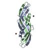
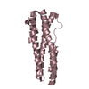
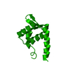
- Assembly
| Deposited unit | 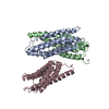
| ||||||||
|---|---|---|---|---|---|---|---|---|---|
| 1 | 
| ||||||||
| 2 | 
| ||||||||
| 3 | 
| ||||||||
| 4 | 
| ||||||||
| 5 | 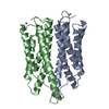
| ||||||||
| Unit cell |
|
-Components
| #1: Protein | Mass: 17956.994 Da / Num. of mol.: 3 / Fragment: Residues 482-655 Source method: isolated from a genetically manipulated source Source: (gene. exp.)  #2: Chemical | ChemComp-PT /   Mass: 195.078 Da / Num. of mol.: 7 / Source method: obtained synthetically / Formula: Pt Mass: 195.078 Da / Num. of mol.: 7 / Source method: obtained synthetically / Formula: Pt#3: Water | ChemComp-HOH / |  Mass: 18.015 Da / Num. of mol.: 130 / Source method: isolated from a natural source / Formula: H2O Mass: 18.015 Da / Num. of mol.: 130 / Source method: isolated from a natural source / Formula: H2O |
|---|
-Experimental details
-Experiment
| Experiment | Method: X-RAY DIFFRACTION / Number of used crystals: 3 |
|---|
- Sample preparation
| Crystal | Density Matthews: 2.82 Å3/Da / Density % sol: 56.44 % |
|---|---|
| Crystal grow | Temperature: 298 K / Method: vapor diffusion, sitting drop / pH: 4.6 Details: 30% PEG 2000 MME, 0.1M Ammonium Acetate, 0.2M Ammonium Sulfate, pH 4.6, VAPOR DIFFUSION, SITTING DROP, temperature 298K |
-Data collection
| Diffraction | Mean temperature: 100 K |
|---|---|
| Diffraction source | Source: SYNCHROTRON / Site: ESRF  / Beamline: ID14-4 / Wavelength: 0.9793 Å / Beamline: ID14-4 / Wavelength: 0.9793 Å |
| Detector | Type: ADSC QUANTUM 4 / Detector: CCD |
| Radiation | Monochromator: 0.9 / Protocol: SINGLE WAVELENGTH / Monochromatic (M) / Laue (L): M / Scattering type: x-ray |
| Radiation wavelength | Wavelength: 0.9793 Å / Relative weight: 1 |
| Reflection | Resolution: 2.5→30 Å / Num. all: 21554 / Num. obs: 21513 / % possible obs: 99.8 % / Rmerge(I) obs: 0.059 / Net I/σ(I): 28.05 |
| Reflection shell | Resolution: 2.5→2.59 Å / Rmerge(I) obs: 0.312 / Mean I/σ(I) obs: 3.576 / Num. unique all: 2120 / % possible all: 100 |
- Processing
| Software |
| ||||||||||||||||||||||||||||||||||||||||||||||||||||||||||||||||||||||||||||||||
|---|---|---|---|---|---|---|---|---|---|---|---|---|---|---|---|---|---|---|---|---|---|---|---|---|---|---|---|---|---|---|---|---|---|---|---|---|---|---|---|---|---|---|---|---|---|---|---|---|---|---|---|---|---|---|---|---|---|---|---|---|---|---|---|---|---|---|---|---|---|---|---|---|---|---|---|---|---|---|---|---|---|
| Refinement | Method to determine structure: SAD / Resolution: 2.5→28.163 Å / Cor.coef. Fo:Fc: 0.928 / Cor.coef. Fo:Fc free: 0.912 / SU B: 10.699 / SU ML: 0.24 / Cross valid method: THROUGHOUT / ESU R: 0.514 / ESU R Free: 0.313 / Stereochemistry target values: MAXIMUM LIKELIHOOD
| ||||||||||||||||||||||||||||||||||||||||||||||||||||||||||||||||||||||||||||||||
| Solvent computation | Ion probe radii: 0.8 Å / Shrinkage radii: 0.8 Å / VDW probe radii: 1.4 Å / Solvent model: BABINET MODEL WITH MASK | ||||||||||||||||||||||||||||||||||||||||||||||||||||||||||||||||||||||||||||||||
| Displacement parameters | Biso mean: 38.414 Å2
| ||||||||||||||||||||||||||||||||||||||||||||||||||||||||||||||||||||||||||||||||
| Refinement step | Cycle: LAST / Resolution: 2.5→28.163 Å
| ||||||||||||||||||||||||||||||||||||||||||||||||||||||||||||||||||||||||||||||||
| Refine LS restraints |
| ||||||||||||||||||||||||||||||||||||||||||||||||||||||||||||||||||||||||||||||||
| LS refinement shell | Resolution: 2.5→2.564 Å / Total num. of bins used: 20 /
|
