 Movie
Movie Controller

[English] 日本語
 Yorodumi
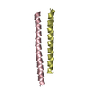
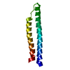
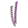
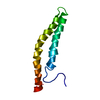
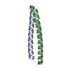
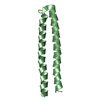
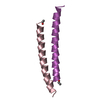
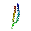
- PDB-1c94: REVERSING THE SEQUENCE OF THE GCN4 LEUCINE ZIPPER DOES NOT AFFECT... -
+ Open data

- Basic information
| Entry | Database: PDB / ID: 1c94 | ||||||
|---|---|---|---|---|---|---|---|
| Title | REVERSING THE SEQUENCE OF THE GCN4 LEUCINE ZIPPER DOES NOT AFFECT ITS FOLD. | ||||||
 Components Components | RETRO-GCN4 LEUCINE ZIPPER | ||||||
 Keywords Keywords | GENE REGULATION / RETRO-COILED COIL / 4-ALPHA-HELIX-BUNDLE / PEPTIDE SYNTHESIS | ||||||
| Method |  X-RAY DIFFRACTION / SYNCHROTRON / Resolution: 2.08 Å X-RAY DIFFRACTION / SYNCHROTRON / Resolution: 2.08 Å | ||||||
 Authors Authors | Mittl, P.R.E. / Deillon, C.A. / Sargent, D. / Liu, N. / Klauser, S. / Thomas, R.M. / Gutte, B. / Gruetter, M.G. | ||||||
 Citation Citation | Journal: Proc.Natl.Acad.Sci.USA / Year: 2000 Title: The retro-GCN4 leucine zipper sequence forms a stable three-dimensional structure. Authors: Mittl, P.R. / Deillon, C. / Sargent, D. / Liu, N. / Klauser, S. / Thomas, R.M. / Gutte, B. / Grutter, M.G. #1: Journal: Science / Year: 1991Title: X-Ray Structure of the GCN4 Leucine Zipper, a Two-Stranded, Parallel Coiled Coil. Authors: O'Shea, E.K. / Klemm, J.D. / Kim, P.S. / Alber, T. #2: Journal: Science / Year: 1993Title: A Switch Between Two-, Three-, and Four-Stranded Coiled Coils in GCN4 Leucine Zipper Mutants. Authors: Harbury, P.B. / Zhang, T. / Kim, P.S. / Alber, T. | ||||||
| History |
|
- Structure visualization
| Structure viewer | Molecule:  MolmilJmol/JSmol MolmilJmol/JSmol |
|---|
- Downloads & links
-Download
| PDBx/mmCIF format | 1c94.cif.gz | 26.7 KB | Display | PDBx/mmCIF format |
|---|---|---|---|---|
| PDB format | pdb1c94.ent.gz | 18.7 KB | Display | PDB format |
| PDBx/mmJSON format | 1c94.json.gz | Tree view | PDBx/mmJSON format | |
| Others |  Other downloads Other downloads |
-Validation report
| Arichive directory | https://data.pdbj.org/pub/pdb/validation_reports/c9/1c94ftp://data.pdbj.org/pub/pdb/validation_reports/c9/1c94 | HTTPS FTP |
|---|
-Related structure data
| Similar structure data |
|---|


-Links
 PDBj
PDBj- Assembly
| Deposited unit | 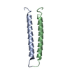
| ||||||||
|---|---|---|---|---|---|---|---|---|---|
| 1 |
| ||||||||
| 2 | 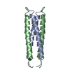
| ||||||||
| Unit cell |
|
-Components
| #1: Protein/peptide | Mass: 4464.196 Da / Num. of mol.: 2 / Source method: obtained synthetically / Details: THIS PEPTIDE WAS CHEMICALLY SYNTHESIZED #2: Water | ChemComp-HOH / |  Mass: 18.015 Da / Num. of mol.: 59 / Source method: isolated from a natural source / Formula: H2O Mass: 18.015 Da / Num. of mol.: 59 / Source method: isolated from a natural source / Formula: H2O |
|---|
-Experimental details
-Experiment
| Experiment | Method: X-RAY DIFFRACTION / Number of used crystals: 1 |
|---|
- Sample preparation
| Crystal | Density Matthews: 1.84 Å3/Da / Density % sol: 33.07 % | |||||||||||||||||||||||||
|---|---|---|---|---|---|---|---|---|---|---|---|---|---|---|---|---|---|---|---|---|---|---|---|---|---|---|
| Crystal grow | Temperature: 295 K / Method: vapor diffusion, hanging drop / pH: 4.8 Details: 25% 2-METHYL-2,4-PENTANEDIOL, 100 MM SODIUM ACETATE, 200 MM SODIUM CHLORIDE., pH 4.8, VAPOR DIFFUSION, HANGING DROP, temperature 295K | |||||||||||||||||||||||||
| Crystal grow | *PLUS Details: drop contained 1:1 mixture of peptide and reservoir solution | |||||||||||||||||||||||||
| Components of the solutions | *PLUS
|
-Data collection
| Diffraction |
| |||||||||||||||
|---|---|---|---|---|---|---|---|---|---|---|---|---|---|---|---|---|
| Diffraction source |
| |||||||||||||||
| Detector |
| |||||||||||||||
| Radiation | Protocol: SINGLE WAVELENGTH / Monochromatic (M) / Laue (L): M / Scattering type: x-ray | |||||||||||||||
| Radiation wavelength | Wavelength: 0.873 Å / Relative weight: 1 | |||||||||||||||
| Reflection | Resolution: 2.08→20 Å / Num. all: 4280 / Num. obs: 3740 / % possible obs: 87.4 % / Redundancy: 2.8 % / Rmerge(I) obs: 0.035 | |||||||||||||||
| Reflection shell | Resolution: 2→2.1 Å / Rmerge(I) obs: 0.11 / % possible all: 80.7 | |||||||||||||||
| Reflection | *PLUS % possible obs: 87.2 % | |||||||||||||||
| Reflection shell | *PLUS % possible obs: 80.7 % |

- Processing
| Software |
| ||||||||||||||||||||||||||||||||||||||||||||||||||||||||||||
|---|---|---|---|---|---|---|---|---|---|---|---|---|---|---|---|---|---|---|---|---|---|---|---|---|---|---|---|---|---|---|---|---|---|---|---|---|---|---|---|---|---|---|---|---|---|---|---|---|---|---|---|---|---|---|---|---|---|---|---|---|---|
| Refinement | Resolution: 2.08→20 Å / σ(F): 0 / σ(I): 0 Stereochemistry target values: CNS-TOPPAR PROTEIN_REP.PARAM CNS-TOPPAR WATER_REP.PARAM Details: PHASE SOLUTION FOUND THROUGH MOLECULAR REPLACEMENT.
| ||||||||||||||||||||||||||||||||||||||||||||||||||||||||||||
| Refinement step | Cycle: LAST / Resolution: 2.08→20 Å
| ||||||||||||||||||||||||||||||||||||||||||||||||||||||||||||
| Refine LS restraints |
|
