 Movie
Movie Controller

[English] 日本語
 Yorodumi
- PDB-1sfl: 1.9A Crystal structure of Staphylococcus aureus type I 3-dehydroq... -
+ Open data

- Basic information
| Entry | Database: PDB / ID: 1sfl | ||||||
|---|---|---|---|---|---|---|---|
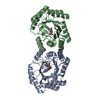
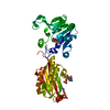
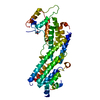
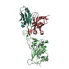
| Title | 1.9A Crystal structure of Staphylococcus aureus type I 3-dehydroquinase, apo form | ||||||
 Components Components | 3-dehydroquinate dehydratase | ||||||
 Keywords Keywords | LYASE / 3-dehydroquinase / enzyme turnover / shikimate pathway / 3-dehydroquinate | ||||||
| Function / homology |  Function and homology information Function and homology information3,4-dihydroxybenzoate biosynthetic process / 3-dehydroquinate dehydratase / 3-dehydroquinate dehydratase activity / chorismate biosynthetic process / aromatic amino acid biosynthetic process / amino acid biosynthetic process Similarity search - Function | ||||||
| Biological species |   Staphylococcus aureus subsp. aureus (bacteria) Staphylococcus aureus subsp. aureus (bacteria) | ||||||
| Method |  X-RAY DIFFRACTION / SYNCHROTRON / MOLECULAR REPLACEMENT / Resolution: 1.9 Å X-RAY DIFFRACTION / SYNCHROTRON / MOLECULAR REPLACEMENT / Resolution: 1.9 Å | ||||||
 Authors Authors | Nichols, C.E. / Lockyer, M. / Hawkins, A.R. / Stammers, D.K. | ||||||
 Citation Citation | Journal: Proteins / Year: 2004 Title: Crystal structures of Staphylococcus aureus type I dehydroquinase from enzyme turnover experiments. Authors: Nichols, C.E. / Lockyer, M. / Hawkins, A.R. / Stammers, D.K. | ||||||
| History |
|
- Structure visualization
| Structure viewer | Molecule: MolmilJmol/JSmol |
|---|
- Downloads & links
-Download
| PDBx/mmCIF format | 1sfl.cif.gz | 112.1 KB | Display | PDBx/mmCIF format |
|---|---|---|---|---|
| PDB format | pdb1sfl.ent.gz | 86.7 KB | Display | PDB format |
| PDBx/mmJSON format | 1sfl.json.gz | Tree view | PDBx/mmJSON format | |
| Others |  Other downloads Other downloads |
-Validation report
| Arichive directory | https://data.pdbj.org/pub/pdb/validation_reports/sf/1sflftp://data.pdbj.org/pub/pdb/validation_reports/sf/1sfl | HTTPS FTP |
|---|
-Related structure data
| Related structure data |  1sfjSC S: Starting model for refinement C: citing same article ( |
|---|---|
| Similar structure data |


-Links
 PDBj
PDBj
- Assembly
| Deposited unit | 
| ||||||||
|---|---|---|---|---|---|---|---|---|---|
| 1 |
| ||||||||
| Unit cell |
|
-Components
| #1: Protein | Mass: 27002.975 Da / Num. of mol.: 2 Source method: isolated from a genetically manipulated source Source: (gene. exp.) Staphylococcus aureus subsp. aureus (bacteria)Species: Staphylococcus aureus / Strain: MRSA252 / Gene: aroD / Plasmid: pRF88 / Species (production host): Escherichia coli / Production host: References: UniProt: Q8NXI0, UniProt: Q6GII7*PLUS, 3-dehydroquinate dehydratase #2: Water | ChemComp-HOH / |  Mass: 18.015 Da / Num. of mol.: 409 / Source method: isolated from a natural source / Formula: H2O Mass: 18.015 Da / Num. of mol.: 409 / Source method: isolated from a natural source / Formula: H2O |
|---|
-Experimental details
-Experiment
| Experiment | Method: X-RAY DIFFRACTION / Number of used crystals: 1 |
|---|
- Sample preparation
| Crystal | Density Matthews: 2.41 Å3/Da / Density % sol: 48.93 % |
|---|---|
| Crystal grow | Temperature: 277 K / pH: 8 Details: PEG 8000, KCl, TRIS, vapour diffusion, temperature 277K |
-Data collection
| Diffraction | Mean temperature: 100 K |
|---|---|
| Diffraction source | Source: SYNCHROTRON / Site: ESRF  / Beamline: ID14-4 / Wavelength: 0.9333 / Wavelength: 0.9333 Å / Beamline: ID14-4 / Wavelength: 0.9333 / Wavelength: 0.9333 Å |
| Detector | Detector: ADSC / Date: Dec 1, 2002 |
| Radiation | Protocol: SINGLE WAVELENGTH / Monochromatic (M) / Laue (L): M / Scattering type: x-ray |
| Radiation wavelength | Wavelength: 0.9333 Å / Relative weight: 1 |
| Reflection | Resolution: 1.9→30 Å / Num. all: 42584 / Num. obs: 40455 / % possible obs: 95 % / Observed criterion σ(I): -1.5 / Redundancy: 10.6 % / Biso Wilson estimate: 26.3 Å2 / Rmerge(I) obs: 0.053 / Net I/σ(I): 37.4 |
| Reflection shell | Resolution: 1.9→1.95 Å / Redundancy: 3.3 % / Rmerge(I) obs: 0.212 / Mean I/σ(I) obs: 5.1 / Num. unique all: 1756 / % possible all: 82.8 |
- Processing
| Software |
| |||||||||||||||||||||||||
|---|---|---|---|---|---|---|---|---|---|---|---|---|---|---|---|---|---|---|---|---|---|---|---|---|---|---|
| Refinement | Method to determine structure: MOLECULAR REPLACEMENT Starting model: PDB ENTRY 1SFJ Resolution: 1.9→30 Å / Cross valid method: THROUGHOUT / σ(I): -1.5 / Stereochemistry target values: Engh and Huber
| |||||||||||||||||||||||||
| Displacement parameters | Biso mean: 49.3 Å2
| |||||||||||||||||||||||||
| Refine analyze |
| |||||||||||||||||||||||||
| Refinement step | Cycle: LAST / Resolution: 1.9→30 Å
| |||||||||||||||||||||||||
| LS refinement shell | Resolution: 1.9→1.95 Å / Rfactor Rfree error: 0.028
|
