 Movie
Movie Controller

[English] 日本語
 Yorodumi
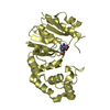
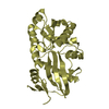
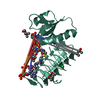
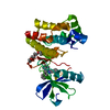
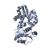
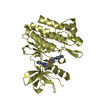
- PDB-1s7j: Crystal structure of phenazine biosynthesis protein PhzF family (... -
+ Open data

- Basic information
| Entry | Database: PDB / ID: 1s7j | ||||||
|---|---|---|---|---|---|---|---|
| Title | Crystal structure of phenazine biosynthesis protein PhzF family (Enterococcus faecalis) | ||||||
 Components Components | phenazine biosynthesis protein PhzF family | ||||||
 Keywords Keywords | BIOSYNTHETIC PROTEIN / phenazine / biosynthesis / bacteria / enterococcus / Structural Genomics / PSI / Protein Structure Initiative / New York SGX Research Center for Structural Genomics / NYSGXRC | ||||||
| Function / homology | Phenazine biosynthesis PhzF protein / Phenazine biosynthesis-like protein / Diaminopimelate Epimerase; Chain A, domain 1 / Diaminopimelate Epimerase; Chain A, domain 1 / isomerase activity / Roll / cytoplasm / Alpha Beta / Phenazine biosynthesis protein PhzF family Function and homology information Function and homology information | ||||||
| Biological species |   Enterococcus faecalis (bacteria) Enterococcus faecalis (bacteria) | ||||||
| Method |  X-RAY DIFFRACTION / MIR / Resolution: 2.3 Å X-RAY DIFFRACTION / MIR / Resolution: 2.3 Å | ||||||
 Authors Authors | Patskovsky, Y. / Almo, S.C. / Burley, S.K. / New York SGX Research Center for Structural Genomics (NYSGXRC) | ||||||
 Citation Citation | Journal: To be Published Title: Crystal structure of phenazine biosynthesis protein from Enterococcus faecalis Authors: Patskovsky, Y. / Almo, S.C. | ||||||
| History |
|
- Structure visualization
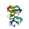
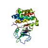
| Structure viewer | Molecule: MolmilJmol/JSmol |
|---|
- Downloads & links
-Download
| PDBx/mmCIF format | 1s7j.cif.gz | 120.1 KB | Display | PDBx/mmCIF format |
|---|---|---|---|---|
| PDB format | pdb1s7j.ent.gz | 93.9 KB | Display | PDB format |
| PDBx/mmJSON format | 1s7j.json.gz | Tree view | PDBx/mmJSON format | |
| Others |  Other downloads Other downloads |
-Validation report
| Arichive directory | https://data.pdbj.org/pub/pdb/validation_reports/s7/1s7jftp://data.pdbj.org/pub/pdb/validation_reports/s7/1s7j | HTTPS FTP |
|---|
-Related structure data
| Similar structure data | |
|---|---|
| Other databases |


-Links
 PDBj
PDBj- Assembly
| Deposited unit | 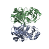
| ||||||||
|---|---|---|---|---|---|---|---|---|---|
| 1 | 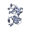
| ||||||||
| 2 | 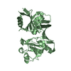
| ||||||||
| Unit cell |
| ||||||||
| Details | probably monomer |
-Components
| #1: Protein | Mass: 29379.416 Da / Num. of mol.: 2 Source method: isolated from a genetically manipulated source Source: (gene. exp.) Enterococcus faecalis (bacteria) / Strain: V583 / Species (production host): Escherichia coli / Production host: #2: Water | ChemComp-HOH / |  Mass: 18.015 Da / Num. of mol.: 348 / Source method: isolated from a natural source / Formula: H2O Mass: 18.015 Da / Num. of mol.: 348 / Source method: isolated from a natural source / Formula: H2O |
|---|
-Experimental details
-Experiment
| Experiment | Method: X-RAY DIFFRACTION / Number of used crystals: 2 |
|---|
- Sample preparation
| Crystal | Density Matthews: 2.38 Å3/Da / Density % sol: 47.84 % |
|---|---|
| Crystal grow | Temperature: 290 K / pH: 7.5 Details: 100 mM HEPES-Na, pH7.5, 27% PEG 3350, 200 mM MgCl2, VAPOR DIFFUSION, SITTING DROP, temperature 290K, pH 7.50 |
-Data collection
| Diffraction | Mean temperature: 100 K |
|---|---|
| Diffraction source | Source: ROTATING ANODE / Type: RIGAKU / Wavelength: 1.5418 |
| Detector | Type: RIGAKU RAXIS IV / Detector: IMAGE PLATE / Date: Dec 8, 2003 / Details: MIRRORS |
| Radiation | Monochromator: MIRRORS / Protocol: SINGLE WAVELENGTH / Monochromatic (M) / Laue (L): M / Scattering type: x-ray |
| Radiation wavelength | Wavelength: 1.5418 Å / Relative weight: 1 |
| Reflection | Resolution: 2.3→20 Å / Num. obs: 25009 / % possible obs: 99.1 % / Observed criterion σ(I): 0 / Redundancy: 18 % / Biso Wilson estimate: 26 Å2 / Rmerge(I) obs: 0.073 / Rsym value: 0.07 / Net I/σ(I): 16.2 |
| Reflection shell | Resolution: 2.3→2.38 Å / Redundancy: 4.1 % / Rmerge(I) obs: 0.176 / Mean I/σ(I) obs: 3.2 / Rsym value: 0.181 / % possible all: 93.9 |
- Processing
| Software |
| ||||||||||||||||||||||||||||||||||||||||||||||||||||||||||||||||||||||||||||||||
|---|---|---|---|---|---|---|---|---|---|---|---|---|---|---|---|---|---|---|---|---|---|---|---|---|---|---|---|---|---|---|---|---|---|---|---|---|---|---|---|---|---|---|---|---|---|---|---|---|---|---|---|---|---|---|---|---|---|---|---|---|---|---|---|---|---|---|---|---|---|---|---|---|---|---|---|---|---|---|---|---|---|
| Refinement | Method to determine structure: MIR / Resolution: 2.3→19.92 Å / Rfactor Rfree error: 0.01 / Data cutoff high absF: 10000 / Data cutoff low absF: 0.1 / Isotropic thermal model: ISOTROPIC / Cross valid method: THROUGHOUT / σ(F): 1 / Stereochemistry target values: ENGH & HUBER Details: The two C-terminal residues were not well-resolved on the maps and were not included in the refinement.
| ||||||||||||||||||||||||||||||||||||||||||||||||||||||||||||||||||||||||||||||||
| Solvent computation | Solvent model: FLAT MODEL / Bsol: 46.8176 Å2 / ksol: 0.339235 e/Å3 | ||||||||||||||||||||||||||||||||||||||||||||||||||||||||||||||||||||||||||||||||
| Displacement parameters | Biso mean: 40.1 Å2
| ||||||||||||||||||||||||||||||||||||||||||||||||||||||||||||||||||||||||||||||||
| Refine analyze |
| ||||||||||||||||||||||||||||||||||||||||||||||||||||||||||||||||||||||||||||||||
| Refinement step | Cycle: LAST / Resolution: 2.3→19.92 Å
| ||||||||||||||||||||||||||||||||||||||||||||||||||||||||||||||||||||||||||||||||
| Refine LS restraints |
| ||||||||||||||||||||||||||||||||||||||||||||||||||||||||||||||||||||||||||||||||
| LS refinement shell | Resolution: 2.3→2.44 Å / Rfactor Rfree error: 0.029 / Total num. of bins used: 6
| ||||||||||||||||||||||||||||||||||||||||||||||||||||||||||||||||||||||||||||||||
| Xplor file |
|
