 Movie
Movie Controller

[English] 日本語
 Yorodumi
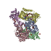
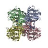
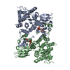
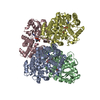
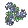
- PDB-1s7g: Structural Basis for the Mechanism and Regulation of Sir2 Enzymes -
+ Open data

- Basic information
| Entry | Database: PDB / ID: 1s7g | ||||||
|---|---|---|---|---|---|---|---|
| Title | Structural Basis for the Mechanism and Regulation of Sir2 Enzymes | ||||||
 Components Components | NAD-dependent deacetylase 2 | ||||||
 Keywords Keywords | TRANSCRIPTION / enzyme / sirtuin / sir2 / NAD+ / ADP-ribose | ||||||
| Function / homology |  Function and homology information Function and homology informationprotein-malonyllysine demalonylase activity / protein-succinyllysine desuccinylase activity / protein acetyllysine N-acetyltransferase / histone deacetylase activity, NAD-dependent / NAD+ binding / zinc ion binding / cytoplasm Similarity search - Function | ||||||
| Biological species |   Archaeoglobus fulgidus (archaea) Archaeoglobus fulgidus (archaea) | ||||||
| Method |  X-RAY DIFFRACTION / SYNCHROTRON / MOLECULAR REPLACEMENT / Resolution: 2.3 Å X-RAY DIFFRACTION / SYNCHROTRON / MOLECULAR REPLACEMENT / Resolution: 2.3 Å | ||||||
 Authors Authors | Avalos, J.L. / Boeke, J.D. / Wolberger, C. | ||||||
 Citation Citation | Journal: Mol.Cell / Year: 2004 Title: Structural basis for the mechanism and regulation of sir2 enzymes Authors: Avalos, J.L. / Boeke, J.D. / Wolberger, C. | ||||||
| History |
|
- Structure visualization
| Structure viewer | Molecule: MolmilJmol/JSmol |
|---|
- Downloads & links
-Download
| PDBx/mmCIF format | 1s7g.cif.gz | 266.9 KB | Display | PDBx/mmCIF format |
|---|---|---|---|---|
| PDB format | pdb1s7g.ent.gz | 211.3 KB | Display | PDB format |
| PDBx/mmJSON format | 1s7g.json.gz | Tree view | PDBx/mmJSON format | |
| Others |  Other downloads Other downloads |
-Validation report
| Arichive directory | https://data.pdbj.org/pub/pdb/validation_reports/s7/1s7gftp://data.pdbj.org/pub/pdb/validation_reports/s7/1s7g | HTTPS FTP |
|---|
-Related structure data
| Related structure data |  1ma3S S: Starting model for refinement |
|---|---|
| Similar structure data |


-Links
 PDBj
PDBj

- Assembly
| Deposited unit | 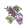
| ||||||||
|---|---|---|---|---|---|---|---|---|---|
| 1 |
| ||||||||
| Unit cell |
|
-Components
-Protein , 1 types, 5 molecules ABCDE
| #1: Protein | Mass: 28537.006 Da / Num. of mol.: 5 Source method: isolated from a genetically manipulated source Source: (gene. exp.) Archaeoglobus fulgidus (archaea) / Gene: NPDA2, AF0112 / Plasmid: pET11a / Production host:  References: UniProt: O30124, Hydrolases; Acting on carbon-nitrogen bonds, other than peptide bonds; In linear amides |
|---|
-Non-polymers , 10 types, 346 molecules 


| #2: Chemical | ChemComp-ZN /  Mass: 65.409 Da / Num. of mol.: 9 / Source method: obtained synthetically / Formula: Zn Mass: 65.409 Da / Num. of mol.: 9 / Source method: obtained synthetically / Formula: Zn#3: Chemical | ChemComp-SO4 /  Mass: 96.063 Da / Num. of mol.: 12 / Source method: obtained synthetically / Formula: SO4 Mass: 96.063 Da / Num. of mol.: 12 / Source method: obtained synthetically / Formula: SO4#4: Chemical |  Mass: 663.425 Da / Num. of mol.: 3 / Source method: obtained synthetically / Formula: C21H27N7O14P2 / Comment: NAD*YM Mass: 663.425 Da / Num. of mol.: 3 / Source method: obtained synthetically / Formula: C21H27N7O14P2 / Comment: NAD*YM#5: Chemical | ChemComp-1PE / |  Mass: 238.278 Da / Num. of mol.: 1 / Source method: obtained synthetically / Formula: C10H22O6 / Comment: precipitant*YM Mass: 238.278 Da / Num. of mol.: 1 / Source method: obtained synthetically / Formula: C10H22O6 / Comment: precipitant*YM#6: Chemical |  Mass: 62.068 Da / Num. of mol.: 3 / Source method: obtained synthetically / Formula: C2H6O2 Mass: 62.068 Da / Num. of mol.: 3 / Source method: obtained synthetically / Formula: C2H6O2#7: Chemical | ChemComp-2PE / |  Mass: 414.488 Da / Num. of mol.: 1 / Source method: obtained synthetically / Formula: C18H38O10 / Comment: precipitant*YM Mass: 414.488 Da / Num. of mol.: 1 / Source method: obtained synthetically / Formula: C18H38O10 / Comment: precipitant*YM#8: Chemical | ChemComp-P6G / |  Mass: 282.331 Da / Num. of mol.: 1 / Source method: obtained synthetically / Formula: C12H26O7 / Comment: precipitant*YM Mass: 282.331 Da / Num. of mol.: 1 / Source method: obtained synthetically / Formula: C12H26O7 / Comment: precipitant*YM#9: Chemical |  Mass: 194.226 Da / Num. of mol.: 2 / Source method: obtained synthetically / Formula: C8H18O5 / Comment: precipitant*YM Mass: 194.226 Da / Num. of mol.: 2 / Source method: obtained synthetically / Formula: C8H18O5 / Comment: precipitant*YM#10: Chemical | ChemComp-APR / |  Mass: 559.316 Da / Num. of mol.: 1 / Source method: obtained synthetically / Formula: C15H23N5O14P2 Mass: 559.316 Da / Num. of mol.: 1 / Source method: obtained synthetically / Formula: C15H23N5O14P2#11: Water | ChemComp-HOH / | Mass: 18.015 Da / Num. of mol.: 313 / Source method: isolated from a natural source / Formula: H2O |
|---|
-Experimental details
-Experiment
| Experiment | Method: X-RAY DIFFRACTION / Number of used crystals: 1 |
|---|
- Sample preparation
| Crystal | Density Matthews: 2.63 Å3/Da / Density % sol: 53.25 % | ||||||||||||||||||||||||||||||||||||||||||
|---|---|---|---|---|---|---|---|---|---|---|---|---|---|---|---|---|---|---|---|---|---|---|---|---|---|---|---|---|---|---|---|---|---|---|---|---|---|---|---|---|---|---|---|
| Crystal grow | Temperature: 293 K / Method: vapor diffusion, hanging drop / pH: 7.4 Details: ammonium sulfate, PEG400, hepes, pH 7.4, VAPOR DIFFUSION, HANGING DROP, temperature 293K | ||||||||||||||||||||||||||||||||||||||||||
| Crystal grow | *PLUS Temperature: 20 ℃ / Method: vapor diffusion, hanging drop | ||||||||||||||||||||||||||||||||||||||||||
| Components of the solutions | *PLUS
|
-Data collection
| Diffraction | Mean temperature: 100 K |
|---|---|
| Diffraction source | Source: SYNCHROTRON / Site: NSLS  / Beamline: X25 / Wavelength: 1.099997 Å / Beamline: X25 / Wavelength: 1.099997 Å |
| Detector | Type: ADSC QUANTUM 4 / Detector: CCD / Date: Apr 16, 2003 |
| Radiation | Monochromator: 2 crystal Si monochromator / Protocol: SINGLE WAVELENGTH / Monochromatic (M) / Laue (L): M / Scattering type: x-ray |
| Radiation wavelength | Wavelength: 1.099997 Å / Relative weight: 1 |
| Reflection | Resolution: 2.3→500 Å / Num. all: 67743 / Num. obs: 67550 / % possible obs: 99.7 % / Observed criterion σ(F): 2 / Observed criterion σ(I): 2 / Redundancy: 9.6 % / Biso Wilson estimate: 30.3 Å2 / Rmerge(I) obs: 0.115 / Rsym value: 0.109 / Net I/σ(I): 15.5 |
| Reflection shell | Resolution: 2.3→2.38 Å / Redundancy: 8.8 % / Rmerge(I) obs: 0.49 / Mean I/σ(I) obs: 4.5 / Num. unique all: 6662 / Rsym value: 0.462 / % possible all: 99 |
| Reflection | *PLUS Num. measured all: 650116 / Rmerge(I) obs: 0.109 |
| Reflection shell | *PLUS % possible obs: 99 % / Num. unique obs: 6662 / Num. measured obs: 58315 / Rmerge(I) obs: 0.462 |
- Processing
| Software |
| ||||||||||||||||||||||||||||||||||||
|---|---|---|---|---|---|---|---|---|---|---|---|---|---|---|---|---|---|---|---|---|---|---|---|---|---|---|---|---|---|---|---|---|---|---|---|---|---|
| Refinement | Method to determine structure: MOLECULAR REPLACEMENT Starting model: 1MA3 Resolution: 2.3→29.78 Å / Rfactor Rfree error: 0.004 / Data cutoff high absF: 1655844.73 / Data cutoff low absF: 0 / Isotropic thermal model: RESTRAINED / Cross valid method: THROUGHOUT / σ(F): 0 / Stereochemistry target values: Engh & Huber
| ||||||||||||||||||||||||||||||||||||
| Solvent computation | Solvent model: FLAT MODEL / Bsol: 28.3174 Å2 / ksol: 0.318351 e/Å3 | ||||||||||||||||||||||||||||||||||||
| Displacement parameters | Biso mean: 41 Å2
| ||||||||||||||||||||||||||||||||||||
| Refine analyze |
| ||||||||||||||||||||||||||||||||||||
| Refinement step | Cycle: LAST / Resolution: 2.3→29.78 Å
| ||||||||||||||||||||||||||||||||||||
| Refine LS restraints |
| ||||||||||||||||||||||||||||||||||||
| LS refinement shell | Resolution: 2.3→2.44 Å / Rfactor Rfree error: 0.013 / Total num. of bins used: 6
| ||||||||||||||||||||||||||||||||||||
| Xplor file |
| ||||||||||||||||||||||||||||||||||||
| Refinement | *PLUS Highest resolution: 2.3 Å / Lowest resolution: 30 Å / Num. reflection obs: 64042 / % reflection Rfree: 5 % / Rfactor Rfree: 0.25 | ||||||||||||||||||||||||||||||||||||
| Solvent computation | *PLUS | ||||||||||||||||||||||||||||||||||||
| Displacement parameters | *PLUS | ||||||||||||||||||||||||||||||||||||
| Refine LS restraints | *PLUS
| ||||||||||||||||||||||||||||||||||||
| LS refinement shell | *PLUS Lowest resolution: 2.38 Å |
