 Movie
Movie Controller

[English] 日本語
 Yorodumi
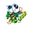
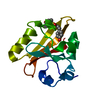
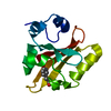
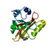
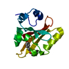
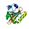
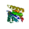
- PDB-1s4s: Reaction Intermediate in the Photocycle of PYP, intermediate occu... -
+ Open data

- Basic information
| Entry | Database: PDB / ID: 1s4s | ||||||
|---|---|---|---|---|---|---|---|
| Title | Reaction Intermediate in the Photocycle of PYP, intermediate occupied between 100 micro-seconds to 5 milli-seconds | ||||||
 Components Components | Photoactive yellow protein | ||||||
 Keywords Keywords | PHOTOSYNTHESIS / reaction intermediate | ||||||
| Function / homology |  Function and homology information Function and homology informationphotoreceptor activity / phototransduction / regulation of DNA-templated transcription / identical protein binding Similarity search - Function | ||||||
| Biological species |  Halorhodospira halophila (bacteria) Halorhodospira halophila (bacteria) | ||||||
| Method |  X-RAY DIFFRACTION / SYNCHROTRON / MOLECULAR REPLACEMENT / Resolution: 1.9 Å X-RAY DIFFRACTION / SYNCHROTRON / MOLECULAR REPLACEMENT / Resolution: 1.9 Å | ||||||
 Authors Authors | Schmidt, M. / Pahl, R. / Srajer, V. / Anderson, S. / Ren, Z. / Ihee, H. / Rajagopal, S. / Moffat, K. | ||||||
 Citation Citation | Journal: Proc.Natl.Acad.Sci.USA / Year: 2004 Title: Protein kinetics: Structures of intermediates and reaction mechanism from time-resolved x-ray data Authors: Schmidt, M. / Pahl, R. / Srajer, V. / Anderson, S. / Ren, Z. / Ihee, H. / Rajagopal, S. / Moffat, K. #1: Journal: Biophys.J. / Year: 2003Title: Application of singular value decomposition to the analysis of time-resolved macromolecular x-ray data Authors: Schmidt, M. / Rajagopal, S. / Ren, Z. / Moffat, K. | ||||||
| History |
|
- Structure visualization
| Structure viewer | Molecule: MolmilJmol/JSmol |
|---|
- Downloads & links
-Download
| PDBx/mmCIF format | 1s4s.cif.gz | 41.2 KB | Display | PDBx/mmCIF format |
|---|---|---|---|---|
| PDB format | pdb1s4s.ent.gz | 28.2 KB | Display | PDB format |
| PDBx/mmJSON format | 1s4s.json.gz | Tree view | PDBx/mmJSON format | |
| Others |  Other downloads Other downloads |
-Validation report
| Arichive directory | https://data.pdbj.org/pub/pdb/validation_reports/s4/1s4sftp://data.pdbj.org/pub/pdb/validation_reports/s4/1s4s | HTTPS FTP |
|---|
-Related structure data
| Related structure data |  1s4rC  2phyS C: citing same article ( S: Starting model for refinement |
|---|---|
| Similar structure data |


-Links
 PDBj
PDBj


- Assembly
| Deposited unit | 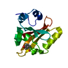
| ||||||||
|---|---|---|---|---|---|---|---|---|---|
| 1 |
| ||||||||
| Unit cell |
|
-Components
| #1: Protein | Mass: 13888.575 Da / Num. of mol.: 1 Source method: isolated from a genetically manipulated source Source: (gene. exp.) Halorhodospira halophila (bacteria) / Production host: |
|---|---|
| #2: Chemical | ChemComp-HC4 /   Mass: 164.158 Da / Num. of mol.: 1 / Source method: obtained synthetically / Formula: C9H8O3 Mass: 164.158 Da / Num. of mol.: 1 / Source method: obtained synthetically / Formula: C9H8O3 |
| #3: Water | ChemComp-HOH /  Mass: 18.015 Da / Num. of mol.: 89 / Source method: isolated from a natural source / Formula: H2O Mass: 18.015 Da / Num. of mol.: 89 / Source method: isolated from a natural source / Formula: H2O |
-Experimental details
-Experiment
| Experiment | Method: X-RAY DIFFRACTION / Number of used crystals: 10 |
|---|
- Sample preparation
| Crystal | Density Matthews: 1.9 Å3/Da / Density % sol: 35.17 % |
|---|---|
| Crystal grow | Temperature: 291 K / Method: vapor diffusion, hanging drop / pH: 7 Details: Ammoniumsulphate, Potassium Phosphate, pH 7.0, VAPOR DIFFUSION, HANGING DROP, temperature 291K |
-Data collection
| Diffraction |
| |||||||||||||||
|---|---|---|---|---|---|---|---|---|---|---|---|---|---|---|---|---|
| Diffraction source |
| |||||||||||||||
| Detector |
| |||||||||||||||
| Radiation |
| |||||||||||||||
| Radiation wavelength |
| |||||||||||||||
| Reflection | Resolution: 1.9→40 Å / Num. all: 8326 / Num. obs: 8326 / % possible obs: 100 % / Observed criterion σ(F): 3 / Observed criterion σ(I): 3 |

- Processing
| Software |
| ||||||||||||||||||||
|---|---|---|---|---|---|---|---|---|---|---|---|---|---|---|---|---|---|---|---|---|---|
| Refinement | Method to determine structure: MOLECULAR REPLACEMENT Starting model: 2phy Resolution: 1.9→15 Å / σ(F): 0 / Stereochemistry target values: Engh & Huber Details: Residue 69 consits of Cys69 and the attached chromophore hydroxy-cinnamic acid. Dihedral of atoms O1 (carbonyl oxygen of chromophore), C1, C2 and C3 was inititally restrained to 0 deg (cis ...Details: Residue 69 consits of Cys69 and the attached chromophore hydroxy-cinnamic acid. Dihedral of atoms O1 (carbonyl oxygen of chromophore), C1, C2 and C3 was inititally restrained to 0 deg (cis planar). The restraint was reset to zero in the last cycle, the angle refined to 22 deg.
| ||||||||||||||||||||
| Refinement step | Cycle: LAST / Resolution: 1.9→15 Å
|
