 Movie
Movie Controller


+ Open data

- Basic information
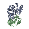
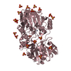
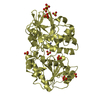
| Entry | Database: PDB / ID: 1s0w | ||||||
|---|---|---|---|---|---|---|---|
| Title | 1b Lactamse/ b Lactamase Inhibitor | ||||||
 Components Components |
| ||||||
 Keywords Keywords | HYDROLASE / PROTEIN-PROTEIN COMPLEX / TEM-1 BETA-LACTAMASE / BETA-LACTAMASE INHIBITOR PROTEIN / BLIP / Israel Structural Proteomics Center / ISPC / Structural Genomics | ||||||
| Function / homology |  Function and homology information Function and homology informationregulation of beta-lactamase activity / Antimicrobial resistance / beta-lactam antibiotic catabolic process / beta-lactamase activity / beta-lactamase / response to antibiotic / extracellular region Similarity search - Function | ||||||
| Biological species |  Streptomyces clavuligerus (bacteria) Streptomyces clavuligerus (bacteria) | ||||||
| Method |  X-RAY DIFFRACTION / MOLECULAR REPLACEMENT / Resolution: 2.3 Å X-RAY DIFFRACTION / MOLECULAR REPLACEMENT / Resolution: 2.3 Å | ||||||
 Authors Authors | Schreiber, G. / Reichman, D. / Israel Structural Proteomics Center (ISPC) | ||||||
 Citation Citation | Journal: Proc.Natl.Acad.Sci.USA / Year: 2005 Title: The modular architecture of protein-protein binding interfaces. Authors: Reichmann, D. / Rahat, O. / Albeck, S. / Meged, R. / Dym, O. / Schreiber, G. | ||||||
| History |
|
- Structure visualization
| Structure viewer | Molecule: MolmilJmol/JSmol |
|---|
- Downloads & links
-Download
| PDBx/mmCIF format | 1s0w.cif.gz | 179.5 KB | Display | PDBx/mmCIF format |
|---|---|---|---|---|
| PDB format | pdb1s0w.ent.gz | 141 KB | Display | PDB format |
| PDBx/mmJSON format | 1s0w.json.gz | Tree view | PDBx/mmJSON format | |
| Others |  Other downloads Other downloads |
-Validation report
| Arichive directory | https://data.pdbj.org/pub/pdb/validation_reports/s0/1s0wftp://data.pdbj.org/pub/pdb/validation_reports/s0/1s0w | HTTPS FTP |
|---|
-Related structure data
| Related structure data |  1xxmC 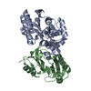 1jtgS S: Starting model for refinement C: citing same article ( |
|---|---|
| Similar structure data | |
| Other databases |


-Links
 PDBj
PDBj


- Assembly
| Deposited unit | 
| ||||||||
|---|---|---|---|---|---|---|---|---|---|
| 1 | 
| ||||||||
| 2 | 
| ||||||||
| Unit cell |
|
-Components
| #1: Protein | Mass: 28941.994 Da / Num. of mol.: 2 Source method: isolated from a genetically manipulated source Source: (gene. exp.) #2: Protein | Mass: 17480.396 Da / Num. of mol.: 2 / Mutation: F1142A Source method: isolated from a genetically manipulated source Source: (gene. exp.) Streptomyces clavuligerus (bacteria) / Production host: #3: Chemical |   Mass: 40.078 Da / Num. of mol.: 2 / Source method: obtained synthetically / Formula: Ca Mass: 40.078 Da / Num. of mol.: 2 / Source method: obtained synthetically / Formula: Ca#4: Water | ChemComp-HOH / |  Mass: 18.015 Da / Num. of mol.: 393 / Source method: isolated from a natural source / Formula: H2O Mass: 18.015 Da / Num. of mol.: 393 / Source method: isolated from a natural source / Formula: H2OHas protein modification | Y | |
|---|
-Experimental details
-Experiment
| Experiment | Method: X-RAY DIFFRACTION / Number of used crystals: 1 |
|---|
- Sample preparation
| Crystal | Density Matthews: 2.46 Å3/Da / Density % sol: 49.93 % | ||||||||||||||||||||||||||||||
|---|---|---|---|---|---|---|---|---|---|---|---|---|---|---|---|---|---|---|---|---|---|---|---|---|---|---|---|---|---|---|---|
| Crystal grow | Temperature: 293 K / Method: microbatch / pH: 7.5 Details: PEG 8000, Dioxane, pH 7.5, Microbatch, temperature 293K | ||||||||||||||||||||||||||||||
| Crystal grow | *PLUS Method: batch method | ||||||||||||||||||||||||||||||
| Components of the solutions | *PLUS
|
-Data collection
| Diffraction | Mean temperature: 120 K |
|---|---|
| Diffraction source | Source: ROTATING ANODE / Type: RIGAKU RU300 / Wavelength: 1.5418 Å |
| Detector | Type: RIGAKU RAXIS IV / Detector: IMAGE PLATE |
| Radiation | Protocol: SINGLE WAVELENGTH / Monochromatic (M) / Laue (L): M / Scattering type: x-ray |
| Radiation wavelength | Wavelength: 1.5418 Å / Relative weight: 1 |
| Reflection | Resolution: 2.3→40 Å / Num. obs: 40572 / % possible obs: 96.9 % / Redundancy: 7 % / Biso Wilson estimate: 27.1 Å2 / Rmerge(I) obs: 0.05 / Rsym value: 0.057 / Net I/σ(I): 22 |
| Reflection shell | Resolution: 2.3→2.38 Å / Redundancy: 5 % / Rmerge(I) obs: 0.189 / Mean I/σ(I) obs: 20.3 / Num. unique all: 3657 / Rsym value: 0.04 / % possible all: 89.9 |
| Reflection | *PLUS Num. obs: 40514 / % possible obs: 97.2 % / Num. measured all: 498302 / Rmerge(I) obs: 0.057 |
| Reflection shell | *PLUS % possible obs: 89.9 % / Rmerge(I) obs: 0.203 / Mean I/σ(I) obs: 6 |
- Processing
| Software |
| ||||||||||||||||||||
|---|---|---|---|---|---|---|---|---|---|---|---|---|---|---|---|---|---|---|---|---|---|
| Refinement | Method to determine structure: MOLECULAR REPLACEMENT Starting model: PDB ENTRY 1jtg Resolution: 2.3→40 Å / σ(F): 0 / σ(I): 0 / Stereochemistry target values: Engh & Huber
| ||||||||||||||||||||
| Refinement step | Cycle: LAST / Resolution: 2.3→40 Å
| ||||||||||||||||||||
| Refine LS restraints |
| ||||||||||||||||||||
| LS refinement shell | Resolution: 2.3→2.32 Å /
| ||||||||||||||||||||
| Refinement | *PLUS Highest resolution: 3 Å / % reflection Rfree: 10 % / Rfactor Rfree: 0.262 / Rfactor Rwork: 0.213 | ||||||||||||||||||||
| Solvent computation | *PLUS | ||||||||||||||||||||
| Displacement parameters | *PLUS |
