 Movie
Movie Controller

[English] 日本語
 Yorodumi
- PDB-1ryy: Acetobacter turbidans alpha-amino acid ester hydrolase Y206A mutant -
+ Open data

- Basic information
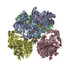
| Entry | Database: PDB / ID: 1ryy | ||||||
|---|---|---|---|---|---|---|---|
| Title | Acetobacter turbidans alpha-amino acid ester hydrolase Y206A mutant | ||||||
 Components Components | alpha-amino acid ester hydrolase | ||||||
 Keywords Keywords | HYDROLASE / alpha/beta hydrolase fold / jellyroll fold | ||||||
| Function / homology |  Function and homology information Function and homology informationalpha-amino-acid esterase / alpha-amino-acid esterase activity / dipeptidyl-peptidase activity Similarity search - Function | ||||||
| Biological species |  Acetobacter pasteurianus (bacteria) Acetobacter pasteurianus (bacteria) | ||||||
| Method |  X-RAY DIFFRACTION / SYNCHROTRON / MOLECULAR REPLACEMENT / Resolution: 2.8 Å X-RAY DIFFRACTION / SYNCHROTRON / MOLECULAR REPLACEMENT / Resolution: 2.8 Å | ||||||
 Authors Authors | Barends, T.R.M. | ||||||
 Citation Citation | Journal: J.Biol.Chem. / Year: 2006 Title: Acetobacter turbidans {alpha}-Amino Acid Ester Hydrolase: HOW A SINGLE MUTATION IMPROVES AN ANTIBIOTIC-PRODUCING ENZYME. Authors: Barends, T.R.M. / Polderman-Tijmes, J.J. / Jekel, P.A. / Williams, C. / Wybenga, G. / Janssen, D.B. / Dijkstra, B.W. | ||||||
| History |
|
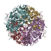
- Structure visualization
| Structure viewer | Molecule: MolmilJmol/JSmol |
|---|
- Downloads & links
-Download
| PDBx/mmCIF format | 1ryy.cif.gz | 815 KB | Display | PDBx/mmCIF format |
|---|---|---|---|---|
| PDB format | pdb1ryy.ent.gz | 663.4 KB | Display | PDB format |
| PDBx/mmJSON format | 1ryy.json.gz | Tree view | PDBx/mmJSON format | |
| Others |  Other downloads Other downloads |
-Validation report
| Arichive directory | https://data.pdbj.org/pub/pdb/validation_reports/ry/1ryyftp://data.pdbj.org/pub/pdb/validation_reports/ry/1ryy | HTTPS FTP |
|---|
-Related structure data


-Links
 PDBj
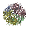
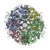
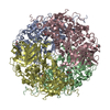
PDBj- Assembly
| Deposited unit | 
| |||||||||||||||||||||||||||||||||||||||||||||||||||||||||||||||||||||||||||||||||||||||||||||||||||||||||||||||||||||||||||||||||||||||||||||||||||||||||||||||||||||||||||||||||||||||||||||||||||||||||||||||||||||||||||||||||||||||||||||
|---|---|---|---|---|---|---|---|---|---|---|---|---|---|---|---|---|---|---|---|---|---|---|---|---|---|---|---|---|---|---|---|---|---|---|---|---|---|---|---|---|---|---|---|---|---|---|---|---|---|---|---|---|---|---|---|---|---|---|---|---|---|---|---|---|---|---|---|---|---|---|---|---|---|---|---|---|---|---|---|---|---|---|---|---|---|---|---|---|---|---|---|---|---|---|---|---|---|---|---|---|---|---|---|---|---|---|---|---|---|---|---|---|---|---|---|---|---|---|---|---|---|---|---|---|---|---|---|---|---|---|---|---|---|---|---|---|---|---|---|---|---|---|---|---|---|---|---|---|---|---|---|---|---|---|---|---|---|---|---|---|---|---|---|---|---|---|---|---|---|---|---|---|---|---|---|---|---|---|---|---|---|---|---|---|---|---|---|---|---|---|---|---|---|---|---|---|---|---|---|---|---|---|---|---|---|---|---|---|---|---|---|---|---|---|---|---|---|---|---|---|---|---|---|---|---|---|---|---|---|---|---|---|---|---|---|---|---|---|
| 1 | 
| |||||||||||||||||||||||||||||||||||||||||||||||||||||||||||||||||||||||||||||||||||||||||||||||||||||||||||||||||||||||||||||||||||||||||||||||||||||||||||||||||||||||||||||||||||||||||||||||||||||||||||||||||||||||||||||||||||||||||||||
| 2 | 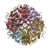
| |||||||||||||||||||||||||||||||||||||||||||||||||||||||||||||||||||||||||||||||||||||||||||||||||||||||||||||||||||||||||||||||||||||||||||||||||||||||||||||||||||||||||||||||||||||||||||||||||||||||||||||||||||||||||||||||||||||||||||||
| Unit cell |
| |||||||||||||||||||||||||||||||||||||||||||||||||||||||||||||||||||||||||||||||||||||||||||||||||||||||||||||||||||||||||||||||||||||||||||||||||||||||||||||||||||||||||||||||||||||||||||||||||||||||||||||||||||||||||||||||||||||||||||||
| Noncrystallographic symmetry (NCS) | NCS domain:
NCS domain segments: Ens-ID: 1 / Refine code: 1
|