 Movie
Movie Controller

[English] 日本語
 Yorodumi
- PDB-2b4k: Acetobacter turbidans alpha-amino acid ester hydrolase complexed ... -
+ Open data

- Basic information
| Entry | Database: PDB / ID: 2b4k | ||||||
|---|---|---|---|---|---|---|---|
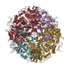
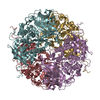
| Title | Acetobacter turbidans alpha-amino acid ester hydrolase complexed with phenylglycine | ||||||
 Components Components | Alpha-amino acid ester hydrolase | ||||||
 Keywords Keywords | HYDROLASE / alpha-beta hydrolase | ||||||
| Function / homology |  Function and homology information Function and homology informationalpha-amino-acid esterase / alpha-amino-acid esterase activity / dipeptidyl-peptidase activity Similarity search - Function | ||||||
| Biological species |  Acetobacter pasteurianus (bacteria) Acetobacter pasteurianus (bacteria) | ||||||
| Method |  X-RAY DIFFRACTION / SYNCHROTRON / MOLECULAR REPLACEMENT / Resolution: 3.3 Å X-RAY DIFFRACTION / SYNCHROTRON / MOLECULAR REPLACEMENT / Resolution: 3.3 Å | ||||||
 Authors Authors | Barends, T.R.M. / Polderman-Tijmes, J.J. / Jekel, P.A. / Williams, C. / Wybenga, G. / Janssen, D.B. / Dijkstra, B.W. | ||||||
 Citation Citation | Journal: J.Biol.Chem. / Year: 2006 Title: Acetobacter turbidans alpha-amino acid ester hydrolase: how a single mutation improves an antibiotic-producing enzyme. Authors: Barends, T.R. / Polderman-Tijmes, J.J. / Jekel, P.A. / Williams, C. / Wybenga, G. / Janssen, D.B. / Dijkstra, B.W. | ||||||
| History |
|
- Structure visualization
| Structure viewer | Molecule: MolmilJmol/JSmol |
|---|
- Downloads & links
-Download
| PDBx/mmCIF format | 2b4k.cif.gz | 422.6 KB | Display | PDBx/mmCIF format |
|---|---|---|---|---|
| PDB format | pdb2b4k.ent.gz | 337.7 KB | Display | PDB format |
| PDBx/mmJSON format | 2b4k.json.gz | Tree view | PDBx/mmJSON format | |
| Others |  Other downloads Other downloads |
-Validation report
| Arichive directory | https://data.pdbj.org/pub/pdb/validation_reports/b4/2b4kftp://data.pdbj.org/pub/pdb/validation_reports/b4/2b4k | HTTPS FTP |
|---|
-Related structure data
| Related structure data |  1nx9SC  1ryyC  2b9vC S: Starting model for refinement C: citing same article ( |
|---|---|
| Similar structure data |


-Links
 PDBj
PDBj- Assembly
| Deposited unit | 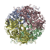
| ||||||||
|---|---|---|---|---|---|---|---|---|---|
| 1 |
| ||||||||
| Unit cell |
|
-Components
| #1: Protein | Mass: 72947.469 Da / Num. of mol.: 4 Source method: isolated from a genetically manipulated source Source: (gene. exp.) Acetobacter pasteurianus (bacteria) / Gene: aehA / Plasmid: pBAD / Production host: #2: Chemical |   Type: D-peptide linking / Mass: 151.163 Da / Num. of mol.: 3 / Source method: obtained synthetically / Formula: C8H9NO2 Type: D-peptide linking / Mass: 151.163 Da / Num. of mol.: 3 / Source method: obtained synthetically / Formula: C8H9NO2#3: Chemical | ChemComp-GOL /   Mass: 92.094 Da / Num. of mol.: 4 / Source method: obtained synthetically / Formula: C3H8O3 Mass: 92.094 Da / Num. of mol.: 4 / Source method: obtained synthetically / Formula: C3H8O3#4: Water | ChemComp-HOH / |  Mass: 18.015 Da / Num. of mol.: 5 / Source method: isolated from a natural source / Formula: H2O Mass: 18.015 Da / Num. of mol.: 5 / Source method: isolated from a natural source / Formula: H2O |
|---|
-Experimental details
-Experiment
| Experiment | Method: X-RAY DIFFRACTION / Number of used crystals: 1 |
|---|
- Sample preparation
| Crystal | Density Matthews: 6.1 Å3/Da / Density % sol: 80 % |
|---|---|
| Crystal grow | Temperature: 293 K / Method: vapor diffusion, hanging drop / pH: 5.6 Details: sodium citrate, PEG4000, phenylglycine, pH 5.6, VAPOR DIFFUSION, HANGING DROP, temperature 293K |
-Data collection
| Diffraction | Mean temperature: 100 K |
|---|---|
| Diffraction source | Source: SYNCHROTRON / Site: ESRF  / Beamline: ID14-4 / Wavelength: 0.934 Å / Beamline: ID14-4 / Wavelength: 0.934 Å |
| Detector | Type: ADSC QUANTUM 4 / Detector: CCD / Date: Jul 1, 2004 |
| Radiation | Monochromator: Double crystal / Protocol: SINGLE WAVELENGTH / Monochromatic (M) / Laue (L): M / Scattering type: x-ray |
| Radiation wavelength | Wavelength: 0.934 Å / Relative weight: 1 |
| Reflection | Resolution: 3→40 Å / Num. all: 121384 / Num. obs: 121384 / % possible obs: 93.2 % / Observed criterion σ(F): 0 / Observed criterion σ(I): 0 / Rmerge(I) obs: 0.186 |
| Reflection shell | Resolution: 3→3.11 Å / Rmerge(I) obs: 0.917 / % possible all: 91.5 |
- Processing
| Software |
| ||||||||||||||||||||||||||||||||||||||||||||||||||
|---|---|---|---|---|---|---|---|---|---|---|---|---|---|---|---|---|---|---|---|---|---|---|---|---|---|---|---|---|---|---|---|---|---|---|---|---|---|---|---|---|---|---|---|---|---|---|---|---|---|---|---|
| Refinement | Method to determine structure: MOLECULAR REPLACEMENT Starting model: pdb entry 1NX9 Resolution: 3.3→15 Å / Cor.coef. Fo:Fc: 0.853 / Cor.coef. Fo:Fc free: 0.813 / SU B: 19.366 / SU ML: 0.33 / Cross valid method: THROUGHOUT / σ(F): 0 / ESU R: 0.669 / ESU R Free: 0.45 / Stereochemistry target values: MAXIMUM LIKELIHOOD
| ||||||||||||||||||||||||||||||||||||||||||||||||||
| Solvent computation | Ion probe radii: 0.8 Å / Shrinkage radii: 0.8 Å / VDW probe radii: 1.4 Å / Solvent model: BABINET MODEL WITH MASK | ||||||||||||||||||||||||||||||||||||||||||||||||||
| Displacement parameters | Biso mean: 19.29 Å2 | ||||||||||||||||||||||||||||||||||||||||||||||||||
| Refinement step | Cycle: LAST / Resolution: 3.3→15 Å
| ||||||||||||||||||||||||||||||||||||||||||||||||||
| Refine LS restraints |
| ||||||||||||||||||||||||||||||||||||||||||||||||||
| LS refinement shell | Resolution: 3.3→3.381 Å / Total num. of bins used: 20 /
|
