 Movie
Movie Controller


+ Open data

- Basic information
| Entry | Database: PDB / ID: 1ryi | |||||||||
|---|---|---|---|---|---|---|---|---|---|---|
| Title | STRUCTURE OF GLYCINE OXIDASE WITH BOUND INHIBITOR GLYCOLATE | |||||||||
 Components Components | GLYCINE OXIDASE | |||||||||
 Keywords Keywords | OXIDOREDUCTASE / FLAVOPROTEIN / OXIDASE / PROTEIN-INHIBITOR COMPLEX | |||||||||
| Function / homology |  Function and homology information Function and homology informationglycine oxidase / glycine oxidase activity / thiamine biosynthetic process / thiamine diphosphate biosynthetic process / response to herbicide / amino acid metabolic process / FAD binding / cytoplasm Similarity search - Function | |||||||||
| Biological species |  | |||||||||
| Method |  X-RAY DIFFRACTION / SYNCHROTRON / MIR / Resolution: 1.8 Å X-RAY DIFFRACTION / SYNCHROTRON / MIR / Resolution: 1.8 Å | |||||||||
 Authors Authors | Moertl, M. / Diederichs, K. / Welte, W. / Pollegioni, L. / Molla, G. / Motteran, L. / Andriolo, G. / Pilone, M.S. | |||||||||
 Citation Citation | Journal: J.Biol.Chem. / Year: 2004 Title: Structure-function correlation in glycine oxidase from Bacillus subtilis Authors: Moertl, M. / Diederichs, K. / Welte, W. / Molla, G. / Motteran, L. / Andriolo, G. / Pilone, M.S. / Pollegioni, L. | |||||||||
| History |
|
- Structure visualization
| Structure viewer | Molecule: MolmilJmol/JSmol |
|---|
- Downloads & links
-Download
| PDBx/mmCIF format | 1ryi.cif.gz | 323.6 KB | Display | PDBx/mmCIF format |
|---|---|---|---|---|
| PDB format | pdb1ryi.ent.gz | 259.3 KB | Display | PDB format |
| PDBx/mmJSON format | 1ryi.json.gz | Tree view | PDBx/mmJSON format | |
| Others |  Other downloads Other downloads |
-Validation report
| Arichive directory | https://data.pdbj.org/pub/pdb/validation_reports/ry/1ryiftp://data.pdbj.org/pub/pdb/validation_reports/ry/1ryi | HTTPS FTP |
|---|
-Related structure data
| Related structure data | |
|---|---|
| Similar structure data |


-Links
 PDBj
PDBj- Assembly
| Deposited unit | 
| ||||||||||
|---|---|---|---|---|---|---|---|---|---|---|---|
| 1 | 
| ||||||||||
| 2 | 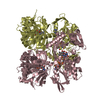
| ||||||||||
| Unit cell |
| ||||||||||
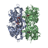
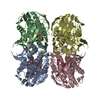
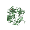
| Details | THE BIOLOGICAL ASSEMBLY IS A TETRAMER. THERE ARE TWO DIFFERENT TETRAMERS IN THE CRYSTAL. THE SECOND PART OF THE FIRST TETRAMER, WICH IS COMPOSED OF CHAIN A AND B AND IT'S SYMMETRY EQUIVALENTS, IS GENERATED BY THE TWO FOLD AXIS x, -y+2, -z+2. THE SECOND PART OF THE SECOND TETRAMER, WICH IS COMPOSED OF CHAIN C AND D AND IT'S SYMMETRY EQUIVALENTS, IS GENERATED BY THE TWO FOLD AXIS -x, y, -z+3/2. |
-Components
| #1: Protein | Mass: 42636.719 Da / Num. of mol.: 4 Source method: isolated from a genetically manipulated source Source: (gene. exp.) #2: Chemical | ChemComp-FAD /   Mass: 785.550 Da / Num. of mol.: 4 / Source method: obtained synthetically / Formula: C27H33N9O15P2 / Comment: FAD*YM Mass: 785.550 Da / Num. of mol.: 4 / Source method: obtained synthetically / Formula: C27H33N9O15P2 / Comment: FAD*YM#3: Chemical | ChemComp-GOA / 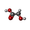  Mass: 76.051 Da / Num. of mol.: 4 / Source method: obtained synthetically / Formula: C2H4O3 Mass: 76.051 Da / Num. of mol.: 4 / Source method: obtained synthetically / Formula: C2H4O3#4: Water | ChemComp-HOH / |  Mass: 18.015 Da / Num. of mol.: 1046 / Source method: isolated from a natural source / Formula: H2O Mass: 18.015 Da / Num. of mol.: 1046 / Source method: isolated from a natural source / Formula: H2O |
|---|
-Experimental details
-Experiment
| Experiment | Method: X-RAY DIFFRACTION / Number of used crystals: 1 |
|---|
- Sample preparation
| Crystal | Density Matthews: 2.46 Å3/Da / Density % sol: 49.5 % |
|---|---|
| Crystal grow | Temperature: 291 K / Method: vapor diffusion, hanging drop / pH: 8.2 Details: 10% w/v PEG 1000, 100 mM Imidazole, 200 mM Ca-Acetate, 30 mM Sodium-Glycolate, pH 8.2, VAPOR DIFFUSION, HANGING DROP, temperature 291K |
-Data collection
| Diffraction | Mean temperature: 100 K |
|---|---|
| Diffraction source | Source: SYNCHROTRON / Site: SRS  / Beamline: PX9.6 / Wavelength: 0.97934 Å / Beamline: PX9.6 / Wavelength: 0.97934 Å |
| Detector | Type: MARRESEARCH / Detector: CCD / Date: Feb 28, 2003 |
| Radiation | Monochromator: Si / Protocol: SINGLE WAVELENGTH / Monochromatic (M) / Laue (L): M / Scattering type: x-ray |
| Radiation wavelength | Wavelength: 0.97934 Å / Relative weight: 1 |
| Reflection | Resolution: 1.8→19.92 Å / Num. all: 159578 / Num. obs: 159578 / Observed criterion σ(F): 0 / Observed criterion σ(I): 0 |
| Reflection shell | Resolution: 1.8→1.9 Å / % possible all: 96.5 |
- Processing
| Software |
| ||||||||||||||||||||||||||||||||||||||||||||||||||||||||||||||||||||||||||||||||||||||||||||||||||||||||||||||||||||||||||||||||||||||||||||||||||||||||||||||||
|---|---|---|---|---|---|---|---|---|---|---|---|---|---|---|---|---|---|---|---|---|---|---|---|---|---|---|---|---|---|---|---|---|---|---|---|---|---|---|---|---|---|---|---|---|---|---|---|---|---|---|---|---|---|---|---|---|---|---|---|---|---|---|---|---|---|---|---|---|---|---|---|---|---|---|---|---|---|---|---|---|---|---|---|---|---|---|---|---|---|---|---|---|---|---|---|---|---|---|---|---|---|---|---|---|---|---|---|---|---|---|---|---|---|---|---|---|---|---|---|---|---|---|---|---|---|---|---|---|---|---|---|---|---|---|---|---|---|---|---|---|---|---|---|---|---|---|---|---|---|---|---|---|---|---|---|---|---|---|---|---|---|
| Refinement | Method to determine structure: MIR / Resolution: 1.8→19.92 Å / Cor.coef. Fo:Fc: 0.963 / Cor.coef. Fo:Fc free: 0.948 / SU B: 2.782 / SU ML: 0.084 / Cross valid method: THROUGHOUT / ESU R: 0.116 / ESU R Free: 0.114 / Stereochemistry target values: MAXIMUM LIKELIHOOD / Details: HYDROGENS HAVE BEEN ADDED IN THE RIDING POSITIONS
| ||||||||||||||||||||||||||||||||||||||||||||||||||||||||||||||||||||||||||||||||||||||||||||||||||||||||||||||||||||||||||||||||||||||||||||||||||||||||||||||||
| Solvent computation | Ion probe radii: 0.8 Å / Shrinkage radii: 0.8 Å / VDW probe radii: 1.4 Å / Solvent model: BABINET MODEL WITH MASK | ||||||||||||||||||||||||||||||||||||||||||||||||||||||||||||||||||||||||||||||||||||||||||||||||||||||||||||||||||||||||||||||||||||||||||||||||||||||||||||||||
| Displacement parameters | Biso mean: 25.85 Å2
| ||||||||||||||||||||||||||||||||||||||||||||||||||||||||||||||||||||||||||||||||||||||||||||||||||||||||||||||||||||||||||||||||||||||||||||||||||||||||||||||||
| Refinement step | Cycle: LAST / Resolution: 1.8→19.92 Å
| ||||||||||||||||||||||||||||||||||||||||||||||||||||||||||||||||||||||||||||||||||||||||||||||||||||||||||||||||||||||||||||||||||||||||||||||||||||||||||||||||
| Refine LS restraints |
| ||||||||||||||||||||||||||||||||||||||||||||||||||||||||||||||||||||||||||||||||||||||||||||||||||||||||||||||||||||||||||||||||||||||||||||||||||||||||||||||||
| LS refinement shell | Resolution: 1.8→1.846 Å / Total num. of bins used: 20 /
|
