 Movie
Movie Controller

[English] 日本語
 Yorodumi
- PDB-1ryb: Crystal Structure of the Chloroplast Group II Intron Splicing Fac... -
+ Open data

- Basic information
| Entry | Database: PDB / ID: 1ryb | ||||||
|---|---|---|---|---|---|---|---|
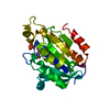
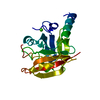
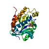
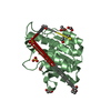
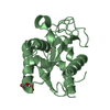
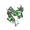
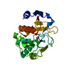
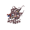
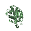
| Title | Crystal Structure of the Chloroplast Group II Intron Splicing Factor CRS2 | ||||||
 Components Components | CRS2 | ||||||
 Keywords Keywords | HYDROLASE / ALPHA-BETA | ||||||
| Function / homology |  Function and homology information Function and homology informationpeptidyl-tRNA hydrolase activity / chloroplast stroma / RNA splicing / mRNA processing / tRNA binding / ribonucleoprotein complex Similarity search - Function | ||||||
| Biological species |  | ||||||
| Method |  X-RAY DIFFRACTION / SYNCHROTRON / MOLECULAR REPLACEMENT / Resolution: 1.7 Å X-RAY DIFFRACTION / SYNCHROTRON / MOLECULAR REPLACEMENT / Resolution: 1.7 Å | ||||||
 Authors Authors | Ostheimer, G.J. / Barkan, A. / Matthews, B.W. | ||||||
 Citation Citation | Journal: J.Mol.Biol. / Year: 2005 Title: Structural analysis of the group II intron splicing factor CRS2 yields insights into its protein and RNA interaction surfaces Authors: Ostheimer, G.J. / Hadjivasiliou, H. / Kloer, D.P. / Barkan, A. / Matthews, B.W. | ||||||
| History |
|
- Structure visualization
| Structure viewer | Molecule: MolmilJmol/JSmol |
|---|
- Downloads & links
-Download
| PDBx/mmCIF format | 1ryb.cif.gz | 53.6 KB | Display | PDBx/mmCIF format |
|---|---|---|---|---|
| PDB format | pdb1ryb.ent.gz | 37 KB | Display | PDB format |
| PDBx/mmJSON format | 1ryb.json.gz | Tree view | PDBx/mmJSON format | |
| Others |  Other downloads Other downloads |
-Validation report
| Summary document | 1ryb_validation.pdf.gz | 418.1 KB | Display | wwPDB validaton report |
|---|---|---|---|---|
| Full document | 1ryb_full_validation.pdf.gz | 419.8 KB | Display | |
| Data in XML | 1ryb_validation.xml.gz | 12.6 KB | Display | |
| Data in CIF | 1ryb_validation.cif.gz | 17 KB | Display | |
| Arichive directory | https://data.pdbj.org/pub/pdb/validation_reports/ry/1rybftp://data.pdbj.org/pub/pdb/validation_reports/ry/1ryb | HTTPS FTP |
-Related structure data
| Related structure data |  1rymC  1rynC  2pthS S: Starting model for refinement C: citing same article ( |
|---|---|
| Similar structure data |

-Links
 PDBj
PDBj- Assembly
| Deposited unit | 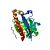
| ||||||||
|---|---|---|---|---|---|---|---|---|---|
| 1 |
| ||||||||
| Unit cell |
|
-Components
| #1: Protein | Mass: 22676.805 Da / Num. of mol.: 1 Source method: isolated from a genetically manipulated source Source: (gene. exp.)  |
|---|---|
| #2: Water | ChemComp-HOH /  Mass: 18.015 Da / Num. of mol.: 164 / Source method: isolated from a natural source / Formula: H2O Mass: 18.015 Da / Num. of mol.: 164 / Source method: isolated from a natural source / Formula: H2O |
-Experimental details
-Experiment
| Experiment | Method: X-RAY DIFFRACTION / Number of used crystals: 1 |
|---|
- Sample preparation
| Crystal | Density Matthews: 1.89 Å3/Da / Density % sol: 34.76 % | |||||||||||||||||||||||||||||||||||
|---|---|---|---|---|---|---|---|---|---|---|---|---|---|---|---|---|---|---|---|---|---|---|---|---|---|---|---|---|---|---|---|---|---|---|---|---|
| Crystal grow | Temperature: 298 K / Method: vapor diffusion, sitting drop / pH: 10.5 Details: PEG 4000, CAPS, sodium chloride, pH 10.5, VAPOR DIFFUSION, SITTING DROP, temperature 298K | |||||||||||||||||||||||||||||||||||
| Crystal grow | *PLUS Method: vapor diffusion, sitting drop | |||||||||||||||||||||||||||||||||||
| Components of the solutions | *PLUS
|
-Data collection
| Diffraction | Mean temperature: 100 K |
|---|---|
| Diffraction source | Source: SYNCHROTRON / Site: SSRL  / Beamline: BL9-1 / Wavelength: 0.773 Å / Beamline: BL9-1 / Wavelength: 0.773 Å |
| Detector | Type: MARRESEARCH / Detector: CCD / Date: Dec 2, 2000 |
| Radiation | Protocol: SINGLE WAVELENGTH / Monochromatic (M) / Laue (L): M / Scattering type: x-ray |
| Radiation wavelength | Wavelength: 0.773 Å / Relative weight: 1 |
| Reflection | Resolution: 1.7→13 Å / Num. all: 20295 / Num. obs: 20208 / % possible obs: 99.6 % / Observed criterion σ(F): 2 / Observed criterion σ(I): 1 / Redundancy: 3.6 % / Biso Wilson estimate: 15.5 Å2 / Rsym value: 0.052 / Net I/σ(I): 8.1 |
| Reflection shell | Resolution: 1.7→1.79 Å / Redundancy: 3.6 % / Mean I/σ(I) obs: 3.9 / Num. unique all: 10361 / Rsym value: 0.194 / % possible all: 99.6 |
| Reflection | *PLUS Lowest resolution: 13.1 Å / Num. obs: 20160 / Rmerge(I) obs: 0.052 |
| Reflection shell | *PLUS % possible obs: 99.6 % / Rmerge(I) obs: 0.194 |
- Processing
| Software |
| |||||||||||||||||||||||||
|---|---|---|---|---|---|---|---|---|---|---|---|---|---|---|---|---|---|---|---|---|---|---|---|---|---|---|
| Refinement | Method to determine structure: MOLECULAR REPLACEMENT Starting model: PDB ENTRY 2PTH Resolution: 1.7→12 Å / Cross valid method: THROUGHOUT / σ(F): 2 / σ(I): 1 / Stereochemistry target values: Engh & Huber
| |||||||||||||||||||||||||
| Refinement step | Cycle: LAST / Resolution: 1.7→12 Å
| |||||||||||||||||||||||||
| Refine LS restraints |
| |||||||||||||||||||||||||
| Refinement | *PLUS Lowest resolution: 13.1 Å / Rfactor Rwork: 0.176 | |||||||||||||||||||||||||
| Solvent computation | *PLUS | |||||||||||||||||||||||||
| Displacement parameters | *PLUS | |||||||||||||||||||||||||
| Refine LS restraints | *PLUS
|
