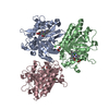
 Movie
Movie Controller

[English] 日本語
 Yorodumi
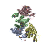
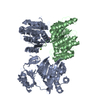
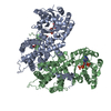
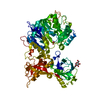
- PDB-1ru3: Crystal Structure of the monomeric acetyl-CoA synthase from Carbo... -
+ Open data

- Basic information
| Entry | Database: PDB / ID: 1ru3 | ||||||
|---|---|---|---|---|---|---|---|
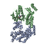
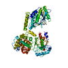
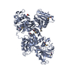
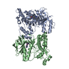
| Title | Crystal Structure of the monomeric acetyl-CoA synthase from Carboxydothermus hydrogenoformans | ||||||
 Components Components | Acetyl-CoA synthase | ||||||
 Keywords Keywords | OXIDOREDUCTASE / Nickel / Cluster A | ||||||
| Function / homology |  Function and homology information Function and homology informationCO-methylating acetyl-CoA synthase / CO-methylating acetyl-CoA synthase activity / anaerobic carbon-monoxide dehydrogenase activity / acetyl-CoA metabolic process / 4 iron, 4 sulfur cluster binding / metal ion binding Similarity search - Function | ||||||
| Biological species |   Carboxydothermus hydrogenoformans (bacteria) Carboxydothermus hydrogenoformans (bacteria) | ||||||
| Method |  X-RAY DIFFRACTION / SYNCHROTRON / MAD / Resolution: 2.2 Å X-RAY DIFFRACTION / SYNCHROTRON / MAD / Resolution: 2.2 Å | ||||||
 Authors Authors | Svetlitchnyi, V. / Dobbek, H. / Meyer-Klaucke, W. / Meins, T. / Thiele, B. / Rmer, P. / Huber, R. / Meyer, O. | ||||||
 Citation Citation | Journal: Proc.Natl.Acad.Sci.USA / Year: 2004 Title: A functional Ni-Ni-[4Fe-4S] cluster in the monomeric acetyl-CoA synthase from Carboxydothermus hydrogenoformans Authors: Svetlitchnyi, V. / Dobbek, H. / Meyer-Klaucke, W. / Meins, T. / Thiele, B. / Rmer, P. / Huber, R. / Meyer, O. | ||||||
| History |
|
- Structure visualization
| Structure viewer | Molecule: MolmilJmol/JSmol |
|---|
- Downloads & links
-Download
| PDBx/mmCIF format | 1ru3.cif.gz | 163.4 KB | Display | PDBx/mmCIF format |
|---|---|---|---|---|
| PDB format | pdb1ru3.ent.gz | 129.4 KB | Display | PDB format |
| PDBx/mmJSON format | 1ru3.json.gz | Tree view | PDBx/mmJSON format | |
| Others |  Other downloads Other downloads |
-Validation report
| Arichive directory | https://data.pdbj.org/pub/pdb/validation_reports/ru/1ru3ftp://data.pdbj.org/pub/pdb/validation_reports/ru/1ru3 | HTTPS FTP |
|---|
-Related structure data
| Similar structure data |
|---|

-Links
 PDBj
PDBj- Assembly
| Deposited unit | 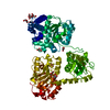
| ||||||||
|---|---|---|---|---|---|---|---|---|---|
| 1 |
| ||||||||
| Unit cell |
|
-Components
| #1: Protein | Mass: 82313.414 Da / Num. of mol.: 1 / Source method: isolated from a natural source Source: (natural) Carboxydothermus hydrogenoformans (bacteria)References: UniProt: P83789 | ||||||||
|---|---|---|---|---|---|---|---|---|---|
| #2: Chemical |   Mass: 58.693 Da / Num. of mol.: 2 / Source method: obtained synthetically / Formula: Ni Mass: 58.693 Da / Num. of mol.: 2 / Source method: obtained synthetically / Formula: Ni#3: Chemical | ChemComp-SF4 / |   Mass: 351.640 Da / Num. of mol.: 1 / Source method: obtained synthetically / Formula: Fe4S4 Mass: 351.640 Da / Num. of mol.: 1 / Source method: obtained synthetically / Formula: Fe4S4#4: Chemical | ChemComp-GOL /   Mass: 92.094 Da / Num. of mol.: 12 / Source method: obtained synthetically / Formula: C3H8O3 Mass: 92.094 Da / Num. of mol.: 12 / Source method: obtained synthetically / Formula: C3H8O3#5: Water | ChemComp-HOH / |  Mass: 18.015 Da / Num. of mol.: 223 / Source method: isolated from a natural source / Formula: H2O Mass: 18.015 Da / Num. of mol.: 223 / Source method: isolated from a natural source / Formula: H2OHas protein modification | Y | |
-Experimental details
-Experiment
| Experiment | Method: X-RAY DIFFRACTION / Number of used crystals: 1 |
|---|
- Sample preparation
| Crystal | Density Matthews: 3.97 Å3/Da / Density % sol: 69.03 % |
|---|---|
| Crystal grow | Temperature: 290 K / Method: vapor diffusion, hanging drop / pH: 6.8 Details: ammonium phosphate, pH 6.8, VAPOR DIFFUSION, HANGING DROP, temperature 290K |
| Crystal grow | *PLUS Temperature: 17 ℃ / Method: vapor diffusion, hanging drop |
| Components of the solutions | *PLUS Conc.: 2 mM / Common name: sodium-dithionite / Details: pH6.8 |
-Data collection
| Diffraction | Mean temperature: 100 K | |||||||||||||||
|---|---|---|---|---|---|---|---|---|---|---|---|---|---|---|---|---|
| Diffraction source | Source: SYNCHROTRON / Site: ESRF  / Beamline: ID14-2 / Wavelength: 1.733, 1.7421, 1.47, 1.05 / Beamline: ID14-2 / Wavelength: 1.733, 1.7421, 1.47, 1.05 | |||||||||||||||
| Detector | Type: ADSC QUANTUM 4 / Detector: CCD / Date: Feb 25, 2002 | |||||||||||||||
| Radiation | Monochromator: Diamond (111), Ge(220) / Protocol: SINGLE WAVELENGTH / Monochromatic (M) / Laue (L): M / Scattering type: x-ray | |||||||||||||||
| Radiation wavelength |
| |||||||||||||||
| Reflection | Resolution: 2.2→20 Å / Num. obs: 64871 / % possible obs: 98.9 % / Observed criterion σ(F): 0 / Observed criterion σ(I): 0 | |||||||||||||||
| Reflection shell | Resolution: 2.2→2.3 Å / % possible all: 97.2 | |||||||||||||||
| Reflection | *PLUS % possible obs: 97.6 % / Num. measured all: 301338 / Rmerge(I) obs: 0.049 | |||||||||||||||
| Reflection shell | *PLUS Mean I/σ(I) obs: 2 |
- Processing
| Software |
| ||||||||||||||||||||
|---|---|---|---|---|---|---|---|---|---|---|---|---|---|---|---|---|---|---|---|---|---|
| Refinement | Method to determine structure: MAD / Resolution: 2.2→20 Å / Cross valid method: THROUGHOUT / σ(F): 0 / Stereochemistry target values: Engh & Huber
| ||||||||||||||||||||
| Refinement step | Cycle: LAST / Resolution: 2.2→20 Å
| ||||||||||||||||||||
| Refinement | *PLUS % reflection Rfree: 5 % | ||||||||||||||||||||
| Solvent computation | *PLUS | ||||||||||||||||||||
| Displacement parameters | *PLUS | ||||||||||||||||||||
| Refine LS restraints | *PLUS
| ||||||||||||||||||||
| LS refinement shell | *PLUS Highest resolution: 2.2 Å / Lowest resolution: 2.3 Å |
