 Movie
Movie Controller

[English] 日本語
 Yorodumi
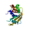
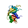
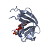
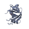
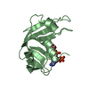
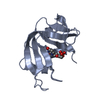
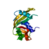
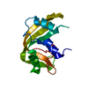
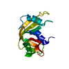
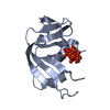
- PDB-1rta: CRYSTAL STRUCTURE DISPOSITION OF THYMIDYLIC ACID TETRAMER IN COMP... -
+ Open data

- Basic information
| Entry | Database: PDB / ID: 1rta | ||||||
|---|---|---|---|---|---|---|---|
| Title | CRYSTAL STRUCTURE DISPOSITION OF THYMIDYLIC ACID TETRAMER IN COMPLEX WITH RIBONUCLEASE A | ||||||
 Components Components |
| ||||||
 Keywords Keywords | HYDROLASE/DNA / PROTEIN-DNA COMPLEX / HYDROLASE-DNA COMPLEX | ||||||
| Function / homology |  Function and homology information Function and homology informationpancreatic ribonuclease / ribonuclease A activity / RNA nuclease activity / nucleic acid binding / lyase activity / defense response to Gram-positive bacterium / extracellular region Similarity search - Function | ||||||
| Biological species |  | ||||||
| Method |  X-RAY DIFFRACTION / Resolution: 2.5 Å X-RAY DIFFRACTION / Resolution: 2.5 Å | ||||||
 Authors Authors | Birdsall, D.L. / McPherson, A. | ||||||
 Citation Citation | Journal: J.Biol.Chem. / Year: 1992 Title: Crystal structure disposition of thymidylic acid tetramer in complex with ribonuclease A. Authors: Birdsall, D.L. / McPherson, A. #1: Journal: Acta Crystallogr.,Sect.B / Year: 1986Title: Comparison of Two Independently Refined Models of Ribonuclease A Authors: Wlodawer, A. / Borkakoti, N. / Moss, D.S. / Howlin, B. #2: Journal: Biochemistry / Year: 1985Title: Nuclear Magnetic Resonance and Neutron Diffraction Studies of the Complex of Ribonuclease A with Uridine Vanadate, a Transition-State Analogue Authors: Borah, B. / Chen, C.-W. / Egan, W. / Miller, M. / Wlodawer, A. / Cohen, J.S. #3: Journal: Proc.Natl.Acad.Sci.USA / Year: 1983Title: Active Site of RNase: Neutron Diffraction Study of a Complex with Uridine Vanadate, a Transition-State Analog Authors: Wlodawer, A. / Miller, M. / Sjolin, L. #4: Journal: Biochemistry / Year: 1983Title: Structure of Ribonuclease A: Results of Joint Neutron and X-Ray Refinement at 2.0 Angstroms Resolution Authors: Wlodawer, A. / Sjolin, L. #5: Journal: J.Biol.Chem. / Year: 1982Title: The Refined Crystal Structure of Ribonuclease A at 2.0 Angstroms Resolution Authors: Wlodawer, A. / Bott, R. / Sjolin, L. #6: Journal: Proc.Natl.Acad.Sci.USA / Year: 1982Title: Hydrogen Exchange in RNase A: Neutron Diffraction Study Authors: Wlodawer, A. / Sjolin, L. #7: Journal: Acta Crystallogr.,Sect.A / Year: 1981Title: Structure of Ribonuclease A: X-Ray and Neutron Refinement Authors: Wlodawer, A. / Bott, R. / Sjolin, L. #8: Journal: Acta Crystallogr.,Sect.A / Year: 1981Title: Joint Refinement of Macromolecular Structures with X-Ray and Neutron Single- Crystal Diffraction Data Authors: Wlodawer, A. / Hendrickson, W.A. #9: Journal: Proc.Natl.Acad.Sci.USA / Year: 1981Title: Orientation of Histidine Residues in RNase A: Neutron Diffraction Study Authors: Wlodawer, A. / Sjolin, L. #10: Journal: Acta Crystallogr.,Sect.B / Year: 1980Title: Studies of Ribonuclease A by X-Ray and Neutron Diffraction Authors: Wlodawer, A. | ||||||
| History |
|
- Structure visualization
| Structure viewer | Molecule: MolmilJmol/JSmol |
|---|
- Downloads & links
-Download
| PDBx/mmCIF format | 1rta.cif.gz | 31.7 KB | Display | PDBx/mmCIF format |
|---|---|---|---|---|
| PDB format | pdb1rta.ent.gz | 25 KB | Display | PDB format |
| PDBx/mmJSON format | 1rta.json.gz | Tree view | PDBx/mmJSON format | |
| Others |  Other downloads Other downloads |
-Validation report
| Summary document | 1rta_validation.pdf.gz | 423.5 KB | Display | wwPDB validaton report |
|---|---|---|---|---|
| Full document | 1rta_full_validation.pdf.gz | 439.5 KB | Display | |
| Data in XML | 1rta_validation.xml.gz | 9.3 KB | Display | |
| Data in CIF | 1rta_validation.cif.gz | 11.3 KB | Display | |
| Arichive directory | https://data.pdbj.org/pub/pdb/validation_reports/rt/1rtaftp://data.pdbj.org/pub/pdb/validation_reports/rt/1rta | HTTPS FTP |
-Related structure data

-Links
 PDBj
PDBj

- Assembly
| Deposited unit | 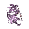
| ||||||||
|---|---|---|---|---|---|---|---|---|---|
| 1 |
| ||||||||
| Unit cell |
| ||||||||
| Atom site foot note | 1: RESIDUES 93 AND 114 ARE CIS PROLINES. |
-Components
| #1: DNA chain | Mass: 1171.814 Da / Num. of mol.: 1 / Source method: obtained synthetically |
|---|---|
| #2: Protein | Mass: 13708.326 Da / Num. of mol.: 1 / Source method: isolated from a natural source / Source: (natural) |
-Experimental details
-Experiment
| Experiment | Method: X-RAY DIFFRACTION |
|---|
- Sample preparation
| Crystal | Density Matthews: 2.47 Å3/Da / Density % sol: 50.12 % |
|---|---|
| Crystal grow | Details: THE COMPLEX WAS FORMED BY DIFFUSION OF DNA INTO THE NATIVE CRYSTALS. |
-Data collection
| Radiation | Protocol: SINGLE WAVELENGTH / Monochromatic (M) / Laue (L): M / Scattering type: x-ray |
|---|---|
| Radiation wavelength | Relative weight: 1 |
| Reflection | *PLUS Highest resolution: 2.5 Å / Num. obs: 4466 / Observed criterion σ(I): 2 |
- Processing
| Software |
| ||||||||||||||||||||||||||||||||||||||||||||||||||||||||||||
|---|---|---|---|---|---|---|---|---|---|---|---|---|---|---|---|---|---|---|---|---|---|---|---|---|---|---|---|---|---|---|---|---|---|---|---|---|---|---|---|---|---|---|---|---|---|---|---|---|---|---|---|---|---|---|---|---|---|---|---|---|---|
| Refinement | Rfactor Rwork: 0.235 / Rfactor obs: 0.235 / Highest resolution: 2.5 Å | ||||||||||||||||||||||||||||||||||||||||||||||||||||||||||||
| Refinement step | Cycle: LAST / Highest resolution: 2.5 Å
| ||||||||||||||||||||||||||||||||||||||||||||||||||||||||||||
| Refine LS restraints |
| ||||||||||||||||||||||||||||||||||||||||||||||||||||||||||||
| Refinement | *PLUS Highest resolution: 2.5 Å / Lowest resolution: 10 Å / Num. reflection obs: 4218 / Rfactor obs: 0.235 | ||||||||||||||||||||||||||||||||||||||||||||||||||||||||||||
| Solvent computation | *PLUS | ||||||||||||||||||||||||||||||||||||||||||||||||||||||||||||
| Displacement parameters | *PLUS | ||||||||||||||||||||||||||||||||||||||||||||||||||||||||||||
| Refine LS restraints | *PLUS Type: x_angle_d / Dev ideal: 5.4 |
