 Movie
Movie Controller

[English] 日本語
 Yorodumi
- PDB-1rir: Crystal structure of meso-tetrasulphonatophenylporphyrin in compl... -
+ Open data

- Basic information
| Entry | Database: PDB / ID: 1rir | ||||||
|---|---|---|---|---|---|---|---|
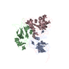
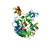
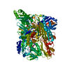
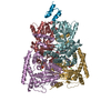
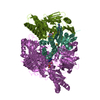
| Title | Crystal structure of meso-tetrasulphonatophenylporphyrin in complex with Peanut lectin. | ||||||
 Components Components | Galactose-binding lectin | ||||||
 Keywords Keywords | SUGAR BINDING PROTEIN | ||||||
| Function / homology |  Function and homology information Function and homology information | ||||||
| Biological species |  | ||||||
| Method |  X-RAY DIFFRACTION / MOLECULAR REPLACEMENT / Resolution: 2.9 Å X-RAY DIFFRACTION / MOLECULAR REPLACEMENT / Resolution: 2.9 Å | ||||||
 Authors Authors | Goel, M. / Kaur, K.J. / Maiya, B.G. / Swamy, M.J. / Salunke, D.M. | ||||||
 Citation Citation | Journal: Biochemistry / Year: 2005 Title: Crystal structures of the PNA-porphyrin complex in the presence and absence of lactose: mapping the conformational changes on lactose binding, interacting surfaces, and supramolecular aggregations. Authors: Goel, M. / Damai, R.S. / Sethi, D.K. / Kaur, K.J. / Maiya, B.G. / Swamy, M.J. / Salunke, D.M. #1: Journal: J.Biol.Chem. / Year: 2001Title: Functional equality in the absence of structural similarity: an added dimension to molecular mimicry. Authors: Goel, M. / Jain, D. / Kaur, K.J. / Kenoth, R. / Maiya, B.G. / Swamy, M.J. / Salunke, D.M. | ||||||
| History |
|
- Structure visualization
| Structure viewer | Molecule: MolmilJmol/JSmol |
|---|
- Downloads & links
-Download
| PDBx/mmCIF format | 1rir.cif.gz | 201.4 KB | Display | PDBx/mmCIF format |
|---|---|---|---|---|
| PDB format | pdb1rir.ent.gz | 166.6 KB | Display | PDB format |
| PDBx/mmJSON format | 1rir.json.gz | Tree view | PDBx/mmJSON format | |
| Others |  Other downloads Other downloads |
-Validation report
| Arichive directory | https://data.pdbj.org/pub/pdb/validation_reports/ri/1rirftp://data.pdbj.org/pub/pdb/validation_reports/ri/1rir | HTTPS FTP |
|---|
-Related structure data
| Related structure data | 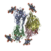 1ritC 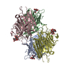 2pelS C: citing same article ( S: Starting model for refinement |
|---|---|
| Similar structure data |


-Links
 PDBj
PDBj

- Assembly
| Deposited unit | 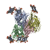
| ||||||||
|---|---|---|---|---|---|---|---|---|---|
| 1 |
| ||||||||
| Unit cell |
|
-Components
| #1: Protein | Mass: 25208.955 Da / Num. of mol.: 4 / Source method: isolated from a natural source / Source: (natural) #2: Chemical | ChemComp-CA /   Mass: 40.078 Da / Num. of mol.: 4 / Source method: obtained synthetically / Formula: Ca Mass: 40.078 Da / Num. of mol.: 4 / Source method: obtained synthetically / Formula: Ca#3: Chemical | ChemComp-MN /   Mass: 54.938 Da / Num. of mol.: 4 / Source method: obtained synthetically / Formula: Mn Mass: 54.938 Da / Num. of mol.: 4 / Source method: obtained synthetically / Formula: Mn#4: Chemical | ChemComp-SFP /   Mass: 939.020 Da / Num. of mol.: 11 / Source method: obtained synthetically / Formula: C44H34N4O12S4 Mass: 939.020 Da / Num. of mol.: 11 / Source method: obtained synthetically / Formula: C44H34N4O12S4#5: Water | ChemComp-HOH / |  Mass: 18.015 Da / Num. of mol.: 61 / Source method: isolated from a natural source / Formula: H2O Mass: 18.015 Da / Num. of mol.: 61 / Source method: isolated from a natural source / Formula: H2O |
|---|
-Experimental details
-Experiment
| Experiment | Method: X-RAY DIFFRACTION / Number of used crystals: 1 |
|---|
- Sample preparation
| Crystal | Density Matthews: 3.7 Å3/Da / Density % sol: 66.8 % |
|---|---|
| Crystal grow | Temperature: 298 K / Method: vapor diffusion, hanging drop / pH: 7.2 Details: ammonium sulphate, Sodium Chloride, Phosphate buffer, pH 7.2, VAPOR DIFFUSION, HANGING DROP, temperature 298.0K |
-Data collection
| Diffraction | Mean temperature: 298 K |
|---|---|
| Diffraction source | Source: ROTATING ANODE / Type: RIGAKU RU200 / Wavelength: 1.5418 Å |
| Detector | Type: MARRESEARCH / Detector: IMAGE PLATE / Date: Jun 11, 2002 |
| Radiation | Monochromator: Graphite / Protocol: SINGLE WAVELENGTH / Monochromatic (M) / Laue (L): M / Scattering type: x-ray |
| Radiation wavelength | Wavelength: 1.5418 Å / Relative weight: 1 |
| Reflection | Resolution: 2.9→100 Å / Num. all: 32142 / Num. obs: 32142 / % possible obs: 91.3 % / Observed criterion σ(F): 0 / Observed criterion σ(I): 0 / Redundancy: 2.3 % / Rmerge(I) obs: 0.1 / Net I/σ(I): 6.5 |
| Reflection shell | Resolution: 2.9→3 Å / Rmerge(I) obs: 0.743 / % possible all: 93.6 |
- Processing
| Software |
| ||||||||||||||||||||
|---|---|---|---|---|---|---|---|---|---|---|---|---|---|---|---|---|---|---|---|---|---|
| Refinement | Method to determine structure: MOLECULAR REPLACEMENT Starting model: 2PEL Resolution: 2.9→100 Å / σ(F): 0 / Stereochemistry target values: Engh & Huber
| ||||||||||||||||||||
| Refinement step | Cycle: LAST / Resolution: 2.9→100 Å
| ||||||||||||||||||||
| LS refinement shell | Resolution: 2.9→2.92 Å / Rfactor Rfree: 0.471 / Rfactor Rwork: 0.436 |
