 Movie
Movie Controller


+ Open data

- Basic information
| Entry | Database: PDB / ID: 1rhy | ||||||
|---|---|---|---|---|---|---|---|
| Title | Crystal structure of Imidazole Glycerol Phosphate Dehydratase | ||||||
 Components Components | Imidazole glycerol phosphate dehydratase | ||||||
 Keywords Keywords | LYASE / dehydratases / histidine biosynthesis / left-handed b-a-b crossover motif / gene duplication | ||||||
| Function / homology |  Function and homology information Function and homology informationimidazoleglycerol-phosphate dehydratase / imidazoleglycerol-phosphate dehydratase activity / L-histidine biosynthetic process Similarity search - Function | ||||||
| Biological species |  Filobasidiella neoformans (Cryptococcus neoformans serotype A) Filobasidiella neoformans (Cryptococcus neoformans serotype A) | ||||||
| Method |  X-RAY DIFFRACTION / SYNCHROTRON / MAD / Resolution: 2.3 Å X-RAY DIFFRACTION / SYNCHROTRON / MAD / Resolution: 2.3 Å | ||||||
 Authors Authors | Sinha, S.C. / Chaudhuri, B.N. / Burgner, J.W. / Yakovleva, G. / Davisson, V.J. / Smith, J.L. | ||||||
 Citation Citation | Journal: J.Biol.Chem. / Year: 2004 Title: Crystal structure of imidazole glycerol-phosphate dehydratase: duplication of an unusual fold Authors: Sinha, S.C. / Chaudhuri, B.N. / Burgner, J.W. / Yakovleva, G. / Davisson, V.J. / Smith, J.L. | ||||||
| History |
|
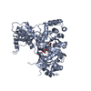
- Structure visualization
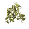
| Structure viewer | Molecule: MolmilJmol/JSmol |
|---|
- Downloads & links
-Download
| PDBx/mmCIF format | 1rhy.cif.gz | 92.3 KB | Display | PDBx/mmCIF format |
|---|---|---|---|---|
| PDB format | pdb1rhy.ent.gz | 70.7 KB | Display | PDB format |
| PDBx/mmJSON format | 1rhy.json.gz | Tree view | PDBx/mmJSON format | |
| Others |  Other downloads Other downloads |
-Validation report
| Arichive directory | https://data.pdbj.org/pub/pdb/validation_reports/rh/1rhyftp://data.pdbj.org/pub/pdb/validation_reports/rh/1rhy | HTTPS FTP |
|---|
-Related structure data
| Similar structure data |
|---|


-Links
 PDBj
PDBj


- Assembly
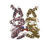
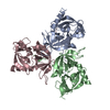
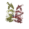
| Deposited unit | 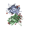
| |||||||||||||||||||||
|---|---|---|---|---|---|---|---|---|---|---|---|---|---|---|---|---|---|---|---|---|---|---|
| 1 | 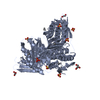
| |||||||||||||||||||||
| 2 | 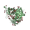
| |||||||||||||||||||||
| Unit cell |
| |||||||||||||||||||||
| Components on special symmetry positions |
| |||||||||||||||||||||
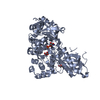
| Details | The biological assembly is a trimer which can be generated from either subunit in the asymmetric unit by the crystallographic three-fold axis, described by the operators x,y,z; y,z,x and z,x,y. |
-Components
-Protein , 1 types, 2 molecules AB
| #1: Protein | Mass: 22008.109 Da / Num. of mol.: 2 Source method: isolated from a genetically manipulated source Source: (gene. exp.) Filobasidiella neoformans (Cryptococcus neoformans serotype A)Gene: HIS3 / Plasmid: pT7 / Production host:  References: UniProt: P40919, UniProt: P0CO22*PLUS, imidazoleglycerol-phosphate dehydratase |
|---|
-Non-polymers , 6 types, 272 molecules 


| #2: Chemical |  Mass: 200.590 Da / Num. of mol.: 2 / Source method: obtained synthetically / Formula: Hg Mass: 200.590 Da / Num. of mol.: 2 / Source method: obtained synthetically / Formula: Hg#3: Chemical | ChemComp-EMC /  Mass: 229.651 Da / Num. of mol.: 4 / Source method: obtained synthetically / Formula: C2H5Hg Mass: 229.651 Da / Num. of mol.: 4 / Source method: obtained synthetically / Formula: C2H5Hg#4: Chemical | ChemComp-SO4 /  Mass: 96.063 Da / Num. of mol.: 5 / Source method: obtained synthetically / Formula: SO4 Mass: 96.063 Da / Num. of mol.: 5 / Source method: obtained synthetically / Formula: SO4#5: Chemical | ChemComp-GOL /  Mass: 92.094 Da / Num. of mol.: 7 / Source method: obtained synthetically / Formula: C3H8O3 Mass: 92.094 Da / Num. of mol.: 7 / Source method: obtained synthetically / Formula: C3H8O3#6: Chemical | ChemComp-ACY /  Mass: 60.052 Da / Num. of mol.: 7 / Source method: obtained synthetically / Formula: C2H4O2 Mass: 60.052 Da / Num. of mol.: 7 / Source method: obtained synthetically / Formula: C2H4O2#7: Water | ChemComp-HOH / | Mass: 18.015 Da / Num. of mol.: 247 / Source method: isolated from a natural source / Formula: H2O |
|---|
-Experimental details
-Experiment
| Experiment | Method: X-RAY DIFFRACTION / Number of used crystals: 1 |
|---|
- Sample preparation
| Crystal | Density Matthews: 2.21 Å3/Da / Density % sol: 43.9 % |
|---|---|
| Crystal grow | Temperature: 293 K / Method: vapor diffusion, hanging drop / pH: 5 Details: ammonium sulfate, sodium acetate,, pH 5.0, VAPOR DIFFUSION, HANGING DROP, temperature 293.0K |
-Data collection
| Diffraction | Mean temperature: 100 K | ||||||||||||
|---|---|---|---|---|---|---|---|---|---|---|---|---|---|
| Diffraction source | Source: SYNCHROTRON / Site: ESRF  / Beamline: BM14 / Wavelength: 1.0086, 1.0078, 0.9150 / Beamline: BM14 / Wavelength: 1.0086, 1.0078, 0.9150 | ||||||||||||
| Detector | Type: MARRESEARCH / Detector: IMAGE PLATE / Date: Sep 15, 1997 | ||||||||||||
| Radiation | Protocol: MAD / Monochromatic (M) / Laue (L): M / Scattering type: x-ray | ||||||||||||
| Radiation wavelength |
| ||||||||||||
| Reflection | Resolution: 2.3→20 Å / Num. all: 17553 / Num. obs: 17324 / % possible obs: 98.7 % / Observed criterion σ(I): 1 / Redundancy: 10 % / Biso Wilson estimate: 31.3 Å2 / Rsym value: 0.066 / Net I/σ(I): 17.8 |
- Processing
| Software |
| ||||||||||||||||||||||||||||||||||||
|---|---|---|---|---|---|---|---|---|---|---|---|---|---|---|---|---|---|---|---|---|---|---|---|---|---|---|---|---|---|---|---|---|---|---|---|---|---|
| Refinement | Method to determine structure: MAD / Resolution: 2.3→19.55 Å / Rfactor Rfree error: 0.008 / Data cutoff high absF: 2905924.67 / Data cutoff low absF: 0 / Isotropic thermal model: RESTRAINED / Cross valid method: THROUGHOUT / σ(F): 0 / σ(I): 0 / Stereochemistry target values: Engh & Huber
| ||||||||||||||||||||||||||||||||||||
| Solvent computation | Solvent model: FLAT MODEL / Bsol: 75.3375 Å2 / ksol: 0.364137 e/Å3 | ||||||||||||||||||||||||||||||||||||
| Displacement parameters | Biso mean: 46.5 Å2 | ||||||||||||||||||||||||||||||||||||
| Refine analyze |
| ||||||||||||||||||||||||||||||||||||
| Refinement step | Cycle: LAST / Resolution: 2.3→19.55 Å
| ||||||||||||||||||||||||||||||||||||
| Refine LS restraints |
| ||||||||||||||||||||||||||||||||||||
| LS refinement shell | Resolution: 2.3→2.44 Å / Rfactor Rfree error: 0.023 / Total num. of bins used: 6
| ||||||||||||||||||||||||||||||||||||
| Xplor file |
|
