 Movie
Movie Controller


+ Open data

- Basic information
| Entry | Database: PDB / ID: 3lgv | ||||||
|---|---|---|---|---|---|---|---|
| Title | H198P mutant of the DegS-deltaPDZ protease | ||||||
 Components Components | Protease degS | ||||||
 Keywords Keywords | HYDROLASE / protease / stress-sensor / HtrA / PDZ OMP / Serine protease | ||||||
| Function / homology |  Function and homology information Function and homology informationpeptidase Do / cellular response to misfolded protein / serine-type peptidase activity / peptidase activity / outer membrane-bounded periplasmic space / serine-type endopeptidase activity / proteolysis / identical protein binding / plasma membrane Similarity search - Function | ||||||
| Biological species |  | ||||||
| Method |  X-RAY DIFFRACTION / MOLECULAR REPLACEMENT / molecular replacement / Resolution: 2.734 Å X-RAY DIFFRACTION / MOLECULAR REPLACEMENT / molecular replacement / Resolution: 2.734 Å | ||||||
 Authors Authors | Sohn, J. / Grant, R.A. / Sauer, R.T. | ||||||
 Citation Citation | Journal: J.Biol.Chem. / Year: 2010 Title: Allostery is an intrinsic property of the protease domain of DegS: implications for enzyme function and evolution. Authors: Sohn, J. / Grant, R.A. / Sauer, R.T. | ||||||
| History |
|
- Structure visualization
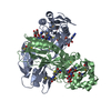
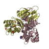
| Structure viewer | Molecule: MolmilJmol/JSmol |
|---|
- Downloads & links
-Download
| PDBx/mmCIF format | 3lgv.cif.gz | 624.1 KB | Display | PDBx/mmCIF format |
|---|---|---|---|---|
| PDB format | pdb3lgv.ent.gz | 524.1 KB | Display | PDB format |
| PDBx/mmJSON format | 3lgv.json.gz | Tree view | PDBx/mmJSON format | |
| Others |  Other downloads Other downloads |
-Validation report
| Arichive directory | https://data.pdbj.org/pub/pdb/validation_reports/lg/3lgvftp://data.pdbj.org/pub/pdb/validation_reports/lg/3lgv | HTTPS FTP |
|---|
-Related structure data
| Related structure data |  3lgiC  3lgtC 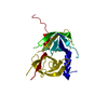 3lguC  3lgwC 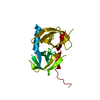 3lgyC  3lh1C  3lh3C C: citing same article ( |
|---|---|
| Similar structure data |


-Links
 PDBj
PDBj
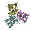
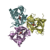
- Assembly
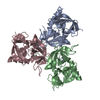
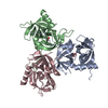
| Deposited unit | 
| ||||||||
|---|---|---|---|---|---|---|---|---|---|
| 1 | 
| ||||||||
| 2 | 
| ||||||||
| 3 | 
| ||||||||
| Unit cell |
|
-Components
| #1: Protein | Mass: 25849.145 Da / Num. of mol.: 9 / Fragment: protease domain / Mutation: h198p Source method: isolated from a genetically manipulated source Source: (gene. exp.) References: UniProt: P0AEE3, Hydrolases; Acting on peptide bonds (peptidases); Serine endopeptidases #2: Water | ChemComp-HOH / |  Mass: 18.015 Da / Num. of mol.: 208 / Source method: isolated from a natural source / Formula: H2O Mass: 18.015 Da / Num. of mol.: 208 / Source method: isolated from a natural source / Formula: H2O |
|---|
-Experimental details
-Experiment
| Experiment | Method: X-RAY DIFFRACTION / Number of used crystals: 1 |
|---|
- Sample preparation
| Crystal | Density Matthews: 2.36 Å3/Da / Density % sol: 47.98 % |
|---|---|
| Crystal grow | Temperature: 300 K / Method: vapor diffusion, sitting drop / pH: 6.5 Details: 50 mM Sodium Cacodylate, 50-125 Sodium Citrate, 10-20 % isopropanol, pH 6.5, VAPOR DIFFUSION, SITTING DROP, temperature 300K |
-Data collection
| Diffraction | Mean temperature: 100 K | ||||||||||||||||||||||||||||||||||||||||||||
|---|---|---|---|---|---|---|---|---|---|---|---|---|---|---|---|---|---|---|---|---|---|---|---|---|---|---|---|---|---|---|---|---|---|---|---|---|---|---|---|---|---|---|---|---|---|
| Diffraction source | Source: ROTATING ANODE / Type: RIGAKU MICROMAX-007 HF / Wavelength: 1.5416 Å | ||||||||||||||||||||||||||||||||||||||||||||
| Detector | Type: RIGAKU RAXIS IV / Detector: IMAGE PLATE / Date: Apr 10, 2009 / Details: Varimax-HF | ||||||||||||||||||||||||||||||||||||||||||||
| Radiation | Monochromator: Varimax-HF / Protocol: SINGLE WAVELENGTH / Monochromatic (M) / Laue (L): M / Scattering type: x-ray | ||||||||||||||||||||||||||||||||||||||||||||
| Radiation wavelength | Wavelength: 1.5416 Å / Relative weight: 1 | ||||||||||||||||||||||||||||||||||||||||||||
| Reflection | Resolution: 2.734→50 Å / Num. obs: 55922 / % possible obs: 94.9 % / Redundancy: 5.6 % / Rmerge(I) obs: 0.097 / Net I/σ(I): 8.8 | ||||||||||||||||||||||||||||||||||||||||||||
| Reflection shell |
|
-Phasing
| Phasing | Method: molecular replacement |
|---|
- Processing
| Software |
| |||||||||||||||||||||||||||||||||||||||||||||||||||||||||||||||||||||||||||||||||||||||||||||||||||||||||||||||||||||||||||||||||||||||||||||||||||
|---|---|---|---|---|---|---|---|---|---|---|---|---|---|---|---|---|---|---|---|---|---|---|---|---|---|---|---|---|---|---|---|---|---|---|---|---|---|---|---|---|---|---|---|---|---|---|---|---|---|---|---|---|---|---|---|---|---|---|---|---|---|---|---|---|---|---|---|---|---|---|---|---|---|---|---|---|---|---|---|---|---|---|---|---|---|---|---|---|---|---|---|---|---|---|---|---|---|---|---|---|---|---|---|---|---|---|---|---|---|---|---|---|---|---|---|---|---|---|---|---|---|---|---|---|---|---|---|---|---|---|---|---|---|---|---|---|---|---|---|---|---|---|---|---|---|---|---|---|
| Refinement | Method to determine structure: MOLECULAR REPLACEMENT / Resolution: 2.734→33.819 Å / Occupancy max: 1 / Occupancy min: 0.5 / SU ML: 0.4 / σ(F): 1.34 / Phase error: 28.69 / Stereochemistry target values: ML Details: To obtain the best possible geometry, this structure was refined with hydrogens whose coordinates were determined by the attached heavy atom. Authors state that although hydrogens cannot be ...Details: To obtain the best possible geometry, this structure was refined with hydrogens whose coordinates were determined by the attached heavy atom. Authors state that although hydrogens cannot be visualized at this resolution, they are present and contribute to scattering. The hydrogens are kept in this entry because independent assessment of many aspects of the geometry, including steric clashes, require their presence. Moreover, removing hydrogen atoms after refinement makes independent assessment of refinement statistics effectively irreproducible.
| |||||||||||||||||||||||||||||||||||||||||||||||||||||||||||||||||||||||||||||||||||||||||||||||||||||||||||||||||||||||||||||||||||||||||||||||||||
| Solvent computation | Shrinkage radii: 0.9 Å / VDW probe radii: 1.11 Å / Solvent model: FLAT BULK SOLVENT MODEL / Bsol: 13.855 Å2 / ksol: 0.321 e/Å3 | |||||||||||||||||||||||||||||||||||||||||||||||||||||||||||||||||||||||||||||||||||||||||||||||||||||||||||||||||||||||||||||||||||||||||||||||||||
| Displacement parameters | Biso mean: 56.528 Å2
| |||||||||||||||||||||||||||||||||||||||||||||||||||||||||||||||||||||||||||||||||||||||||||||||||||||||||||||||||||||||||||||||||||||||||||||||||||
| Refinement step | Cycle: LAST / Resolution: 2.734→33.819 Å
| |||||||||||||||||||||||||||||||||||||||||||||||||||||||||||||||||||||||||||||||||||||||||||||||||||||||||||||||||||||||||||||||||||||||||||||||||||
| Refine LS restraints |
| |||||||||||||||||||||||||||||||||||||||||||||||||||||||||||||||||||||||||||||||||||||||||||||||||||||||||||||||||||||||||||||||||||||||||||||||||||
| LS refinement shell |
|
