 Movie
Movie Controller

[English] 日本語
 Yorodumi
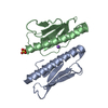
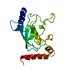
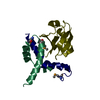
- PDB-1r9k: Representative solution structure of the catalytic domain of SopE2 -
+ Open data

- Basic information
| Entry | Database: PDB / ID: 1r9k | ||||||
|---|---|---|---|---|---|---|---|
| Title | Representative solution structure of the catalytic domain of SopE2 | ||||||
 Components Components | TypeIII-secreted protein effector: invasion-associated protein | ||||||
 Keywords Keywords | CELL INVASION / Salmonella / invasion / type III / GEF / SopE | ||||||
| Function / homology |  Function and homology information Function and homology informationguanyl-nucleotide exchange factor activity / GTPase activator activity / actin cytoskeleton organization / extracellular region Similarity search - Function | ||||||
| Biological species |  Salmonella typhimurium (bacteria) Salmonella typhimurium (bacteria) | ||||||
| Method | SOLUTION NMR / simulated annealing | ||||||
 Authors Authors | Williams, C. / Galyov, E.E. / Bagby, S. | ||||||
 Citation Citation | Journal: Biochemistry / Year: 2004 Title: Solution Structure, Backbone Dynamics, and Interaction with Cdc42 of Salmonella Guanine Nucleotide Exchange Factor SopE2(,). Authors: Williams, C. / Galyov, E.E. / Bagby, S. #1: Journal: To be Published / Year: 2003Title: Biochemical and structural analysis of Salmonella and Burkholderia virulence proteins Authors: Williams, C. #2: Journal: J.BIOMOL.NMR / Year: 2003Title: Assignment of the 1H,13C and 15N resonances of the catalytic domain of guanine nucelotide exchange factor SopE2 from Salmonella dublin Authors: Williams, C. / Galyov, E.E. / Bagby, S. | ||||||
| History |
|
- Structure visualization
| Structure viewer | Molecule: MolmilJmol/JSmol |
|---|
- Downloads & links
-Download
| PDBx/mmCIF format | 1r9k.cif.gz | 67.6 KB | Display | PDBx/mmCIF format |
|---|---|---|---|---|
| PDB format | pdb1r9k.ent.gz | 51.4 KB | Display | PDB format |
| PDBx/mmJSON format | 1r9k.json.gz | Tree view | PDBx/mmJSON format | |
| Others |  Other downloads Other downloads |
-Validation report
| Arichive directory | https://data.pdbj.org/pub/pdb/validation_reports/r9/1r9kftp://data.pdbj.org/pub/pdb/validation_reports/r9/1r9k | HTTPS FTP |
|---|
-Related structure data
| Related structure data | 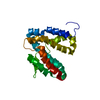 1r6eC C: citing same article ( |
|---|---|
| Similar structure data | |
| Other databases |
|


-Links
 PDBj
PDBj- Assembly
| Deposited unit | 
| |||||||||
|---|---|---|---|---|---|---|---|---|---|---|
| 1 |
| |||||||||
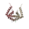
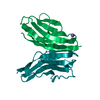
| NMR ensembles |
|
-Components
| #1: Protein | Mass: 18585.338 Da / Num. of mol.: 1 / Fragment: SopE2 GEF domain Source method: isolated from a genetically manipulated source Source: (gene. exp.) Salmonella typhimurium (bacteria) / Strain: LT2 / Gene: sopE2 / Plasmid: pGEX-2T / Species (production host): Escherichia coli / Production host: |
|---|
-Experimental details
-Experiment
| Experiment | Method: SOLUTION NMR | ||||||||||||||||||||||||
|---|---|---|---|---|---|---|---|---|---|---|---|---|---|---|---|---|---|---|---|---|---|---|---|---|---|
| NMR experiment |
| ||||||||||||||||||||||||
| NMR details | Text: The structure was determined using triple-resonance NMR spectroscopy. |
- Sample preparation
| Details |
| ||||||||||||||||||||
|---|---|---|---|---|---|---|---|---|---|---|---|---|---|---|---|---|---|---|---|---|---|
| Sample conditions |
|
-NMR measurement
| Radiation | Protocol: SINGLE WAVELENGTH / Monochromatic (M) / Laue (L): M / Scattering type: x-ray |
|---|---|
| Radiation wavelength | Relative weight: 1 |
| NMR spectrometer | Type: Varian INOVA / Manufacturer: Varian / Model: INOVA / Field strength: 600 MHz |
- Processing
| NMR software |
| ||||||||||||||||||||
|---|---|---|---|---|---|---|---|---|---|---|---|---|---|---|---|---|---|---|---|---|---|
| Refinement | Method: simulated annealing / Software ordinal: 1 Details: he structures are based on a total of 3065 restraints, 2682 are NOE-derived distance constraints, 249 dihedral angle restraints,134 distance restraints from hydrogen bonds. | ||||||||||||||||||||
| NMR representative | Selection criteria: closest to the average | ||||||||||||||||||||
| NMR ensemble | Conformer selection criteria: structures with acceptable covalent geometry,structures with the least restraint violations,structures with the lowest energy Conformers calculated total number: 100 / Conformers submitted total number: 1 |
 NMRPipe