 Movie
Movie Controller

[English] 日本語
 Yorodumi
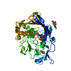
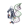
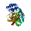
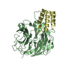
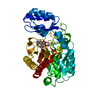
- PDB-1r9f: Crystal structure of p19 complexed with 19-bp small interfering RNA -
+ Open data

- Basic information
| Entry | Database: PDB / ID: 1r9f | ||||||
|---|---|---|---|---|---|---|---|
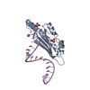
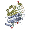
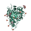
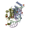
| Title | Crystal structure of p19 complexed with 19-bp small interfering RNA | ||||||
 Components Components |
| ||||||
 Keywords Keywords | Viral protein/RNA / Protein-RNA complex / Dimer / double helix / Viral protein-RNA COMPLEX | ||||||
| Function / homology |  Function and homology information Function and homology informationvirion component / symbiont-mediated suppression of host innate immune response / RNA binding Similarity search - Function | ||||||
| Biological species |  Tomato bushy stunt virus Tomato bushy stunt virus | ||||||
| Method |  X-RAY DIFFRACTION / SYNCHROTRON / MAD / Resolution: 1.85 Å X-RAY DIFFRACTION / SYNCHROTRON / MAD / Resolution: 1.85 Å | ||||||
 Authors Authors | Ye, K. / Malinina, L. / Patel, D.J. | ||||||
 Citation Citation | Journal: Nature / Year: 2003 Title: Recognition of small interfering RNA by a viral suppressor of RNA Authors: Ye, K. / Malinina, L. / Patel, D.J. | ||||||
| History |
|
- Structure visualization
| Structure viewer | Molecule: MolmilJmol/JSmol |
|---|
- Downloads & links
-Download
| PDBx/mmCIF format | 1r9f.cif.gz | 64.3 KB | Display | PDBx/mmCIF format |
|---|---|---|---|---|
| PDB format | pdb1r9f.ent.gz | 42.8 KB | Display | PDB format |
| PDBx/mmJSON format | 1r9f.json.gz | Tree view | PDBx/mmJSON format | |
| Others |  Other downloads Other downloads |
-Validation report
| Arichive directory | https://data.pdbj.org/pub/pdb/validation_reports/r9/1r9fftp://data.pdbj.org/pub/pdb/validation_reports/r9/1r9f | HTTPS FTP |
|---|
-Related structure data
| Similar structure data |
|---|
-Links
 PDBj
PDBj


- Assembly
| Deposited unit | 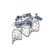
| ||||||||
|---|---|---|---|---|---|---|---|---|---|
| 1 | 
| ||||||||
| Unit cell |
| ||||||||
| Components on special symmetry positions |
| ||||||||
| Details | The biological assemble is a p19 dimer in complex with one siRNA duplex generated from the protein monomer and half RNA (19-bp duplex in half occupancy) in the asymmetric unit by the operation: Y+2/3,X-2/3,-Z+1/3. The 2-fold symmetry operation generates another overlapping half RNA in opposite orientation, compensating the lack of symmetry in RNA duplex itself. |
-Components
| #1: RNA chain | Mass: 6690.004 Da / Num. of mol.: 1 / Source method: obtained synthetically / Details: 5'-OH and 3'-OH | ||||
|---|---|---|---|---|---|
| #2: RNA chain | Mass: 6666.964 Da / Num. of mol.: 1 / Source method: obtained synthetically / Details: 5'-OH and 3'-OH | ||||
| #3: Protein | Mass: 15751.966 Da / Num. of mol.: 1 / Fragment: Residues 27-158 / Mutation: L144M, L147M Source method: isolated from a genetically manipulated source Source: (gene. exp.) Tomato bushy stunt virus / Genus: Tombusvirus / Gene: p19 / Plasmid: pET28a / Species (production host): Escherichia coli / Production host:  | ||||
| #4: Chemical |   Mass: 96.063 Da / Num. of mol.: 2 / Source method: obtained synthetically / Formula: SO4 Mass: 96.063 Da / Num. of mol.: 2 / Source method: obtained synthetically / Formula: SO4#5: Water | ChemComp-HOH / |  Mass: 18.015 Da / Num. of mol.: 70 / Source method: isolated from a natural source / Formula: H2O Mass: 18.015 Da / Num. of mol.: 70 / Source method: isolated from a natural source / Formula: H2OHas protein modification | Y | |
-Experimental details
-Experiment
| Experiment | Method: X-RAY DIFFRACTION / Number of used crystals: 1 |
|---|
- Sample preparation
| Crystal | Density Matthews: 2.05 Å3/Da / Density % sol: 39.86 % | |||||||||||||||||||||||||||||||||||||||||||||||||||||||||||||||
|---|---|---|---|---|---|---|---|---|---|---|---|---|---|---|---|---|---|---|---|---|---|---|---|---|---|---|---|---|---|---|---|---|---|---|---|---|---|---|---|---|---|---|---|---|---|---|---|---|---|---|---|---|---|---|---|---|---|---|---|---|---|---|---|---|
| Crystal grow | Temperature: 293 K / pH: 7.5 Details: ammonium sulfate, potassium chloride, HEPES-NaOH, dithiothreitol, pH 7.5, VAPOR DIFFUSION, HANGING DROP, temperature 293K, pH 7.50 | |||||||||||||||||||||||||||||||||||||||||||||||||||||||||||||||
| Components of the solutions |
| |||||||||||||||||||||||||||||||||||||||||||||||||||||||||||||||
| Crystal grow | *PLUS Temperature: 20 ℃ / pH: 7.6 / Method: vapor diffusion, hanging drop | |||||||||||||||||||||||||||||||||||||||||||||||||||||||||||||||
| Components of the solutions | *PLUS
|
-Data collection
| Diffraction | Mean temperature: 100 K | ||||||||||||
|---|---|---|---|---|---|---|---|---|---|---|---|---|---|
| Diffraction source | Source: SYNCHROTRON / Site: APS  / Beamline: 14-ID-B / Wavelength: 0.9791,0.9789,0.9562 / Beamline: 14-ID-B / Wavelength: 0.9791,0.9789,0.9562 | ||||||||||||
| Detector | Type: MARRESEARCH / Detector: CCD / Date: Jul 18, 2003 | ||||||||||||
| Radiation | Monochromator: DIAMOND (111) DOUBLE-CRYSTAL MONOCHROMATOR / Protocol: MAD / Monochromatic (M) / Laue (L): M / Scattering type: x-ray | ||||||||||||
| Radiation wavelength |
| ||||||||||||
| Reflection | Resolution: 1.85→50 Å / Num. obs: 39408 / % possible obs: 99.1 % / Observed criterion σ(I): -3 / Redundancy: 9.5 % / Biso Wilson estimate: 21.8 Å2 / Rmerge(I) obs: 0.06 / Net I/σ(I): 44.8 | ||||||||||||
| Reflection shell | Resolution: 1.85→1.92 Å / Rmerge(I) obs: 0.725 / Mean I/σ(I) obs: 2 / % possible all: 93.2 | ||||||||||||
| Reflection | *PLUS Highest resolution: 1.85 Å / Lowest resolution: 50 Å / Num. obs: 20413 / % possible obs: 99.11 % / Num. measured all: 194486 / Rmerge(I) obs: 0.06 | ||||||||||||
| Reflection shell | *PLUS % possible obs: 93.2 % / Num. unique obs: 1872 / Mean I/σ(I) obs: 2 |
- Processing
| Software |
| ||||||||||||||||||||||||||||||||||||||||||||||||||||||||||||||||||||||||||||||||
|---|---|---|---|---|---|---|---|---|---|---|---|---|---|---|---|---|---|---|---|---|---|---|---|---|---|---|---|---|---|---|---|---|---|---|---|---|---|---|---|---|---|---|---|---|---|---|---|---|---|---|---|---|---|---|---|---|---|---|---|---|---|---|---|---|---|---|---|---|---|---|---|---|---|---|---|---|---|---|---|---|---|
| Refinement | Method to determine structure: MAD / Resolution: 1.85→14.86 Å / Rfactor Rfree error: 0.005 / Data cutoff low absF: 0 / Isotropic thermal model: RESTRAINED / Cross valid method: THROUGHOUT / σ(F): 0 / Stereochemistry target values: ENGH & HUBER
| ||||||||||||||||||||||||||||||||||||||||||||||||||||||||||||||||||||||||||||||||
| Solvent computation | Solvent model: FLAT MODEL / Bsol: 44.85 Å2 / ksol: 0.35 e/Å3 | ||||||||||||||||||||||||||||||||||||||||||||||||||||||||||||||||||||||||||||||||
| Displacement parameters | Biso mean: 46.4 Å2
| ||||||||||||||||||||||||||||||||||||||||||||||||||||||||||||||||||||||||||||||||
| Refine analyze |
| ||||||||||||||||||||||||||||||||||||||||||||||||||||||||||||||||||||||||||||||||
| Refinement step | Cycle: LAST / Resolution: 1.85→14.86 Å
| ||||||||||||||||||||||||||||||||||||||||||||||||||||||||||||||||||||||||||||||||
| Refine LS restraints |
| ||||||||||||||||||||||||||||||||||||||||||||||||||||||||||||||||||||||||||||||||
| LS refinement shell | Resolution: 1.86→1.97 Å / Rfactor Rfree error: 0.024 / Total num. of bins used: 6
| ||||||||||||||||||||||||||||||||||||||||||||||||||||||||||||||||||||||||||||||||
| Xplor file |
| ||||||||||||||||||||||||||||||||||||||||||||||||||||||||||||||||||||||||||||||||
| Refinement | *PLUS Lowest resolution: 15 Å / % reflection Rfree: 5 % / Rfactor Rfree: 0.236 / Rfactor Rwork: 0.214 | ||||||||||||||||||||||||||||||||||||||||||||||||||||||||||||||||||||||||||||||||
| Solvent computation | *PLUS | ||||||||||||||||||||||||||||||||||||||||||||||||||||||||||||||||||||||||||||||||
| Displacement parameters | *PLUS | ||||||||||||||||||||||||||||||||||||||||||||||||||||||||||||||||||||||||||||||||
| Refine LS restraints | *PLUS
|
