 Movie
Movie Controller


+ Open data

- Basic information
| Entry | Database: PDB / ID: 1dym | |||||||||
|---|---|---|---|---|---|---|---|---|---|---|
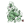
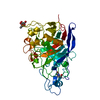
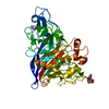
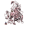
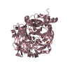
| Title | Humicola insolens Endocellulase Cel7B (EG 1) E197A Mutant | |||||||||
 Components Components | ENDOGLUCANASE I | |||||||||
 Keywords Keywords | HYDROLASE / ENDOGLUCANASE / CELLULOSE DEGRADATION / GLYCOSIDASE / GLYCOPROTEIN / GLYCOSYNTHASE / GLYCOSIDE HYDROLASE FAMILY 7 | |||||||||
| Function / homology |  Function and homology information Function and homology informationcellulase / cellulase activity / cellulose catabolic process / extracellular region Similarity search - Function | |||||||||
| Biological species |  HUMICOLA INSOLENS (fungus) HUMICOLA INSOLENS (fungus) | |||||||||
| Method |  X-RAY DIFFRACTION / SYNCHROTRON / MOLECULAR REPLACEMENT / Resolution: 1.75 Å X-RAY DIFFRACTION / SYNCHROTRON / MOLECULAR REPLACEMENT / Resolution: 1.75 Å | |||||||||
 Authors Authors | Davies, G.J. / Moraz, O. / Driguez, H. / Schulein, M. | |||||||||
 Citation Citation | Journal: Biochem.J. / Year: 1998 Title: Crystal structure of the family 7 endoglucanase I (Cel7B) from Humicola insolens at 2.2 A resolution and identification of the catalytic nucleophile by trapping of the covalent glycosyl-enzyme intermediate. Authors: MacKenzie, L.F. / Sulzenbacher, G. / Divne, C. / Jones, T.A. / Woldike, H.F. / Schulein, M. / Withers, S.G. / Davies, G.J. #1: Journal: J.Biotechnol. / Year: 1997Title: Oligosaccharide Specificity of a Family 7 Endoglucanase: Insertion of Potential Sugar-Binding Subsites Authors: Davies, G.J. / Ducros, V. / Lewis, R.J. / Borchert, T.V. / Schulein, M. | |||||||||
| History |
| |||||||||
| Remark 700 | SHEET DETERMINATION METHOD: PART OF THE SHEET STRUCTURE OF THIS MOLECULE IS BIFURCATED. ORDER TO ... SHEET DETERMINATION METHOD: PART OF THE SHEET STRUCTURE OF THIS MOLECULE IS BIFURCATED. ORDER TO REPRESENT THIS FEATURE IN THE SHEET RECORDS BELOW, TWO SHEETS ARE DEFINED. STRANDS 1, 2, AND 3 OF SHEETS A AND A1 ARE IDENTICAL. |
- Structure visualization
| Structure viewer | Molecule: MolmilJmol/JSmol |
|---|
- Downloads & links
-Download
| PDBx/mmCIF format | 1dym.cif.gz | 106.1 KB | Display | PDBx/mmCIF format |
|---|---|---|---|---|
| PDB format | pdb1dym.ent.gz | 79.8 KB | Display | PDB format |
| PDBx/mmJSON format | 1dym.json.gz | Tree view | PDBx/mmJSON format | |
| Others |  Other downloads Other downloads |
-Validation report
| Arichive directory | https://data.pdbj.org/pub/pdb/validation_reports/dy/1dymftp://data.pdbj.org/pub/pdb/validation_reports/dy/1dym | HTTPS FTP |
|---|
-Related structure data
| Related structure data |  2a39SC S: Starting model for refinement C: citing same article ( |
|---|---|
| Similar structure data |


-Links
 PDBj
PDBj- Assembly
| Deposited unit | 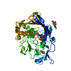
| ||||||||
|---|---|---|---|---|---|---|---|---|---|
| 1 |
| ||||||||
| Unit cell |
|
-Components
| #1: Protein | Mass: 44552.109 Da / Num. of mol.: 1 / Mutation: YES Source method: isolated from a genetically manipulated source Details: PYROGLUTAMATE POST-TRANSLATIONAL MODIFICATION AT RESIDUE 1, N-LINKED N-ACETYLGLUCOSAMINE ON RESIDUE ASNS 89 AND 247 Source: (gene. exp.) HUMICOLA INSOLENS (fungus) / Production host: | ||||
|---|---|---|---|---|---|
| #2: Sugar |   Type: D-saccharide, beta linking / Mass: 221.208 Da / Num. of mol.: 2 Type: D-saccharide, beta linking / Mass: 221.208 Da / Num. of mol.: 2Source method: isolated from a genetically manipulated source Formula: C8H15NO6 #3: Water | ChemComp-HOH / |  Mass: 18.015 Da / Num. of mol.: 595 / Source method: isolated from a natural source / Formula: H2O Mass: 18.015 Da / Num. of mol.: 595 / Source method: isolated from a natural source / Formula: H2OHas protein modification | Y | |
-Experimental details
-Experiment
| Experiment | Method: X-RAY DIFFRACTION / Number of used crystals: 1 |
|---|
- Sample preparation
| Crystal | Density Matthews: 2.43 Å3/Da / Density % sol: 49.45 % |
|---|---|
| Crystal grow | pH: 8 / Details: 20-30% PEG 4K, 20 MM TRIS-HCL PH 8.0 |
-Data collection
| Diffraction | Mean temperature: 100 K |
|---|---|
| Diffraction source | Source: SYNCHROTRON / Site: EMBL/DESY, HAMBURG  / Beamline: X11 / Wavelength: 0.9 / Beamline: X11 / Wavelength: 0.9 |
| Detector | Type: MARRESEARCH / Detector: IMAGE PLATE / Date: Oct 15, 1998 |
| Radiation | Protocol: SINGLE WAVELENGTH / Monochromatic (M) / Laue (L): M / Scattering type: x-ray |
| Radiation wavelength | Wavelength: 0.9 Å / Relative weight: 1 |
| Reflection | Resolution: 1.75→20 Å / Num. obs: 43135 / % possible obs: 100 % / Redundancy: 4.9 % / Biso Wilson estimate: 16 Å2 / Rmerge(I) obs: 0.076 / Rsym value: 0.076 / Net I/σ(I): 19.7 |
| Reflection shell | Resolution: 1.75→1.81 Å / Redundancy: 4.8 % / Rmerge(I) obs: 0.45 / Mean I/σ(I) obs: 3.3 / Rsym value: 0.45 / % possible all: 100 |
- Processing
| Software |
| ||||||||||||||||||||||||||||||||||||||||||||||||||||||||||||||||||||||||||||||||||||
|---|---|---|---|---|---|---|---|---|---|---|---|---|---|---|---|---|---|---|---|---|---|---|---|---|---|---|---|---|---|---|---|---|---|---|---|---|---|---|---|---|---|---|---|---|---|---|---|---|---|---|---|---|---|---|---|---|---|---|---|---|---|---|---|---|---|---|---|---|---|---|---|---|---|---|---|---|---|---|---|---|---|---|---|---|---|
| Refinement | Method to determine structure: MOLECULAR REPLACEMENT Starting model: PDB ENTRY 2A39 NATIVE STRUCTURE Resolution: 1.75→20 Å / Cross valid method: THROUGHOUT / σ(F): 0
| ||||||||||||||||||||||||||||||||||||||||||||||||||||||||||||||||||||||||||||||||||||
| Refinement step | Cycle: LAST / Resolution: 1.75→20 Å
| ||||||||||||||||||||||||||||||||||||||||||||||||||||||||||||||||||||||||||||||||||||
| Refine LS restraints |
|
