 Movie
Movie Controller

[English] 日本語
 Yorodumi
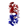
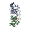
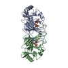
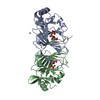
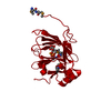
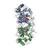
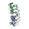
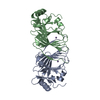
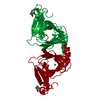
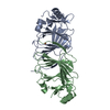
- PDB-1qxj: Crystal structure of native phosphoglucose isomerase from Pyrococ... -
+ Open data

- Basic information
| Entry | Database: PDB / ID: 1qxj | ||||||
|---|---|---|---|---|---|---|---|
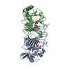
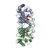
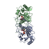
| Title | Crystal structure of native phosphoglucose isomerase from Pyrococcus furiosus | ||||||
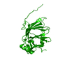
 Components Components | Glucose-6-phosphate isomerase | ||||||
 Keywords Keywords | ISOMERASE / phosphoglucose isomerase / cupin fold / Pyrococcus furiosus / hyperthermophile / extremophile / aldose-ketose isomerase | ||||||
| Function / homology |  Function and homology information Function and homology informationglucose-6-phosphate isomerase / glucose-6-phosphate isomerase activity / gluconeogenesis / glycolytic process / iron ion binding / cytoplasm Similarity search - Function | ||||||
| Biological species |   Pyrococcus furiosus (archaea) Pyrococcus furiosus (archaea) | ||||||
| Method |  X-RAY DIFFRACTION / MOLECULAR REPLACEMENT / Resolution: 1.8 Å X-RAY DIFFRACTION / MOLECULAR REPLACEMENT / Resolution: 1.8 Å | ||||||
 Authors Authors | Swan, M.K. / Solomons, J.T.G. / Beeson, C.C. / Hansen, T. / Schonheit, P. / Davies, C. | ||||||
 Citation Citation | Journal: J.Biol.Chem. / Year: 2003 Title: Structural evidence for a hydride transfer mechanism of catalysis in phosphoglucose isomerase from Pyrococcus furiosus Authors: Swan, M.K. / Solomons, J.T.G. / Beeson, C.C. / Hansen, T. / Schonheit, P. / Davies, C. #1: Journal: Protein Pept.Lett. / Year: 2003Title: Crystallization and preliminary X-ray diffraction analysis of phosphoglucose isomerase from Pyrococcus furiosus Authors: Swan, M.K. / Hansen, T. / Schonheit, P. / Davies, C. | ||||||
| History |
|
- Structure visualization
| Structure viewer | Molecule: MolmilJmol/JSmol |
|---|
- Downloads & links
-Download
| PDBx/mmCIF format | 1qxj.cif.gz | 92.4 KB | Display | PDBx/mmCIF format |
|---|---|---|---|---|
| PDB format | pdb1qxj.ent.gz | 69.7 KB | Display | PDB format |
| PDBx/mmJSON format | 1qxj.json.gz | Tree view | PDBx/mmJSON format | |
| Others |  Other downloads Other downloads |
-Validation report
| Arichive directory | https://data.pdbj.org/pub/pdb/validation_reports/qx/1qxjftp://data.pdbj.org/pub/pdb/validation_reports/qx/1qxj | HTTPS FTP |
|---|
-Related structure data
| Related structure data |  1qxrC  1qy4C  1plz C: citing same article ( S: Starting model for refinement |
|---|---|
| Similar structure data |

-Links
 PDBj
PDBj

- Assembly
| Deposited unit | 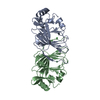
| ||||||||
|---|---|---|---|---|---|---|---|---|---|
| 1 |
| ||||||||
| Unit cell |
|
-Components
| #1: Protein | Mass: 21505.436 Da / Num. of mol.: 2 Source method: isolated from a genetically manipulated source Source: (gene. exp.) Pyrococcus furiosus (archaea) / Gene: PGIA OR PF0196 / Plasmid: pet17b / Species (production host): Escherichia coli / Production host:  #2: Chemical |   Mass: 58.693 Da / Num. of mol.: 2 / Source method: obtained synthetically / Formula: Ni Mass: 58.693 Da / Num. of mol.: 2 / Source method: obtained synthetically / Formula: Ni#3: Water | ChemComp-HOH / |  Mass: 18.015 Da / Num. of mol.: 249 / Source method: isolated from a natural source / Formula: H2O Mass: 18.015 Da / Num. of mol.: 249 / Source method: isolated from a natural source / Formula: H2O |
|---|
-Experimental details
-Experiment
| Experiment | Method: X-RAY DIFFRACTION / Number of used crystals: 1 |
|---|
- Sample preparation
| Crystal | Density Matthews: 2.3 Å3/Da / Density % sol: 45 % | ||||||||||||||||||||||||
|---|---|---|---|---|---|---|---|---|---|---|---|---|---|---|---|---|---|---|---|---|---|---|---|---|---|
| Crystal grow | Temperature: 295 K / Method: vapor diffusion, hanging drop / pH: 5.6 Details: 25% PEG 4000, 0.2M ammonium acetate, 0.1 M sodium acetate pH 5.6, VAPOR DIFFUSION, HANGING DROP, temperature 295K | ||||||||||||||||||||||||
| Crystal grow | *PLUS Method: vapor diffusion, hanging drop | ||||||||||||||||||||||||
| Components of the solutions | *PLUS
|
-Data collection
| Diffraction | Mean temperature: 100 K |
|---|---|
| Diffraction source | Source: ROTATING ANODE / Type: RIGAKU RUH3R / Wavelength: 1.5418 Å |
| Detector | Type: RIGAKU RAXIS IV / Detector: IMAGE PLATE / Date: Jul 4, 2002 / Details: mirrors |
| Radiation | Monochromator: OSMIC MIRRORS / Protocol: SINGLE WAVELENGTH / Monochromatic (M) / Laue (L): M / Scattering type: x-ray |
| Radiation wavelength | Wavelength: 1.5418 Å / Relative weight: 1 |
| Reflection | Resolution: 1.8→36.7 Å / Num. obs: 34845 / % possible obs: 92.1 % / Observed criterion σ(F): 0 / Observed criterion σ(I): 0 / Redundancy: 3.74 % / Biso Wilson estimate: 20.6 Å2 / Rmerge(I) obs: 0.065 / Net I/σ(I): 18.5 |
| Reflection shell | Resolution: 1.8→1.86 Å / Redundancy: 3.8 % / Rmerge(I) obs: 0.324 / Mean I/σ(I) obs: 2.7 / Num. unique all: 3464 / % possible all: 83.4 |
| Reflection | *PLUS Num. measured all: 130434 |
| Reflection shell | *PLUS % possible obs: 83.4 % |
- Processing
| Software |
| |||||||||||||||||||||||||||||||||||||||||||||||||||||||||||||||||||||||||||
|---|---|---|---|---|---|---|---|---|---|---|---|---|---|---|---|---|---|---|---|---|---|---|---|---|---|---|---|---|---|---|---|---|---|---|---|---|---|---|---|---|---|---|---|---|---|---|---|---|---|---|---|---|---|---|---|---|---|---|---|---|---|---|---|---|---|---|---|---|---|---|---|---|---|---|---|---|
| Refinement | Method to determine structure: MOLECULAR REPLACEMENT Starting model: PDB ENTRY 1PLZ 1plz Resolution: 1.8→15 Å / Cor.coef. Fo:Fc: 0.953 / Cor.coef. Fo:Fc free: 0.924 / SU B: 4.437 / SU ML: 0.136 / Cross valid method: THROUGHOUT / σ(F): 0 / ESU R: 0.154 / ESU R Free: 0.15 / Stereochemistry target values: MAXIMUM LIKELIHOOD
| |||||||||||||||||||||||||||||||||||||||||||||||||||||||||||||||||||||||||||
| Solvent computation | Ion probe radii: 0.8 Å / Shrinkage radii: 0.8 Å / VDW probe radii: 1.4 Å / Solvent model: BABINET MODEL WITH MASK | |||||||||||||||||||||||||||||||||||||||||||||||||||||||||||||||||||||||||||
| Displacement parameters | Biso mean: 25.085 Å2
| |||||||||||||||||||||||||||||||||||||||||||||||||||||||||||||||||||||||||||
| Refinement step | Cycle: LAST / Resolution: 1.8→15 Å
| |||||||||||||||||||||||||||||||||||||||||||||||||||||||||||||||||||||||||||
| Refine LS restraints |
| |||||||||||||||||||||||||||||||||||||||||||||||||||||||||||||||||||||||||||
| LS refinement shell | Resolution: 1.8→1.846 Å / Total num. of bins used: 20 /
| |||||||||||||||||||||||||||||||||||||||||||||||||||||||||||||||||||||||||||
| Software | *PLUS Version: 5 / Classification: refinement | |||||||||||||||||||||||||||||||||||||||||||||||||||||||||||||||||||||||||||
| Refinement | *PLUS Highest resolution: 1.8 Å / Rfactor obs: 0.197 / Rfactor Rfree: 0.249 / Rfactor Rwork: 0.194 | |||||||||||||||||||||||||||||||||||||||||||||||||||||||||||||||||||||||||||
| Solvent computation | *PLUS | |||||||||||||||||||||||||||||||||||||||||||||||||||||||||||||||||||||||||||
| Displacement parameters | *PLUS | |||||||||||||||||||||||||||||||||||||||||||||||||||||||||||||||||||||||||||
| Refine LS restraints | *PLUS
|
