 Movie
Movie Controller

[English] 日本語
 Yorodumi
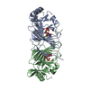
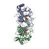
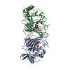
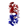
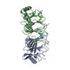
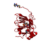
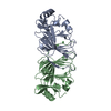
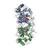
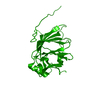
- PDB-4lum: The crystal structure of the P132V mutant of Pyrococcus furiosus ... -
+ Open data

- Basic information
| Entry | Database: PDB / ID: 4lum | ||||||
|---|---|---|---|---|---|---|---|
| Title | The crystal structure of the P132V mutant of Pyrococcus furiosus phosphoglucose isomerase in complex with manganese and fructose-6- phosphate. | ||||||
 Components Components | Glucose-6-phosphate isomerase | ||||||
 Keywords Keywords | ISOMERASE / Cupin Fold / Glucose 6-phosphate and Fructose 6-phosphate binding protein | ||||||
| Function / homology |  Function and homology information Function and homology informationglucose-6-phosphate isomerase / glucose-6-phosphate isomerase activity / gluconeogenesis / glycolytic process / iron ion binding / cytoplasm Similarity search - Function | ||||||
| Biological species |   Pyrococcus furiosus (archaea) Pyrococcus furiosus (archaea) | ||||||
| Method |  X-RAY DIFFRACTION / SYNCHROTRON / MOLECULAR REPLACEMENT / Resolution: 1.79 Å X-RAY DIFFRACTION / SYNCHROTRON / MOLECULAR REPLACEMENT / Resolution: 1.79 Å | ||||||
 Authors Authors | Baker, P.J. / Almourfi, F.M. | ||||||
 Citation Citation | Journal: To be Published Title: Correlated mutation analysis as a tool for smart library design to improve protein performance. Authors: Baker, P.J. / Almourfi, F.M. / Raedts, J. / Joosten, H.-J. / Hendriks, S. / Kengen, S.W.M. / Hage, W.R. / Schaap, P.J. / Sedelnikova, S.E. / Van der Oost, J. | ||||||
| History |
|
- Structure visualization
| Structure viewer | Molecule: MolmilJmol/JSmol |
|---|
- Downloads & links
-Download
| PDBx/mmCIF format | 4lum.cif.gz | 171.5 KB | Display | PDBx/mmCIF format |
|---|---|---|---|---|
| PDB format | pdb4lum.ent.gz | 136.2 KB | Display | PDB format |
| PDBx/mmJSON format | 4lum.json.gz | Tree view | PDBx/mmJSON format | |
| Others |  Other downloads Other downloads |
-Validation report
| Arichive directory | https://data.pdbj.org/pub/pdb/validation_reports/lu/4lumftp://data.pdbj.org/pub/pdb/validation_reports/lu/4lum | HTTPS FTP |
|---|
-Related structure data
| Related structure data | 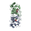 4ltaC 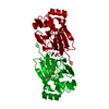 4lukC 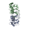 4lulC  1x7nS C: citing same article ( S: Starting model for refinement |
|---|---|
| Similar structure data |


-Links
 PDBj
PDBj

- Assembly
| Deposited unit | 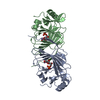
| ||||||||
|---|---|---|---|---|---|---|---|---|---|
| 1 |
| ||||||||
| Unit cell |
| ||||||||
| Details | The asymmetric unit contains the biological dimer. |
-Components
| #1: Protein | Mass: 21638.648 Da / Num. of mol.: 2 / Mutation: P132V Source method: isolated from a genetically manipulated source Source: (gene. exp.) Pyrococcus furiosus (archaea) / Strain: ATCC 43587 / DSM 3638 / JCM 8422 / Vc1 / Gene: pgiA, PF0196 / Plasmid: pET24d / Production host:  #2: Chemical |   Mass: 54.938 Da / Num. of mol.: 2 / Source method: obtained synthetically / Formula: Mn Mass: 54.938 Da / Num. of mol.: 2 / Source method: obtained synthetically / Formula: Mn#3: Chemical | 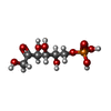  Mass: 260.136 Da / Num. of mol.: 2 / Source method: obtained synthetically / Formula: C6H13O9P Mass: 260.136 Da / Num. of mol.: 2 / Source method: obtained synthetically / Formula: C6H13O9P#4: Water | ChemComp-HOH / |  Mass: 18.015 Da / Num. of mol.: 218 / Source method: isolated from a natural source / Formula: H2O Mass: 18.015 Da / Num. of mol.: 218 / Source method: isolated from a natural source / Formula: H2O |
|---|
-Experimental details
-Experiment
| Experiment | Method: X-RAY DIFFRACTION / Number of used crystals: 1 |
|---|
- Sample preparation
| Crystal | Density Matthews: 2.2 Å3/Da / Density % sol: 44.12 % |
|---|---|
| Crystal grow | Temperature: 290 K / Method: vapor diffusion, sitting drop / pH: 6.5 Details: 0.2 M sodium nitrate, 0.1 M Bis Tris Propane PH6.5, 20% PEG4000, VAPOR DIFFUSION, SITTING DROP, temperature 290K |
-Data collection
| Diffraction | Mean temperature: 100 K |
|---|---|
| Diffraction source | Source: SYNCHROTRON / Site: Diamond  / Beamline: I24 / Wavelength: 0.9686 Å / Beamline: I24 / Wavelength: 0.9686 Å |
| Detector | Type: DECTRIS PILATUS 6M / Detector: PIXEL / Date: Nov 4, 2011 / Details: Diamond I03 |
| Radiation | Monochromator: Diamond I03 / Protocol: SINGLE WAVELENGTH / Monochromatic (M) / Laue (L): M / Scattering type: x-ray |
| Radiation wavelength | Wavelength: 0.9686 Å / Relative weight: 1 |
| Reflection | Resolution: 1.79→18.49 Å / Num. all: 33074 / Num. obs: 33074 / % possible obs: 95.7 % / Observed criterion σ(F): 0 / Observed criterion σ(I): 0 / Redundancy: 1.8 % / Rmerge(I) obs: 0.086 / Net I/σ(I): 4.3 |
| Reflection shell | Resolution: 1.79→1.84 Å / Redundancy: 1.8 % / Rmerge(I) obs: 0.349 / Mean I/σ(I) obs: 2.2 / Num. unique all: 2386 / % possible all: 94 |
- Processing
| Software |
| ||||||||||||||||||||||||||||||||||||||||||||||||||||||||||||||||||||||
|---|---|---|---|---|---|---|---|---|---|---|---|---|---|---|---|---|---|---|---|---|---|---|---|---|---|---|---|---|---|---|---|---|---|---|---|---|---|---|---|---|---|---|---|---|---|---|---|---|---|---|---|---|---|---|---|---|---|---|---|---|---|---|---|---|---|---|---|---|---|---|---|
| Refinement | Method to determine structure: MOLECULAR REPLACEMENT Starting model: PDB ENTRY 1X7N Resolution: 1.79→18.49 Å / Cor.coef. Fo:Fc: 0.961 / Cor.coef. Fo:Fc free: 0.943 / SU B: 6.138 / SU ML: 0.088 / Cross valid method: THROUGHOUT / ESU R: 0.451 / ESU R Free: 0.13 / Stereochemistry target values: MAXIMUM LIKELIHOOD / Details: HYDROGENS HAVE BEEN ADDED IN THE RIDING POSITIONS
| ||||||||||||||||||||||||||||||||||||||||||||||||||||||||||||||||||||||
| Solvent computation | Ion probe radii: 0.8 Å / Shrinkage radii: 0.8 Å / VDW probe radii: 1.4 Å / Solvent model: MASK | ||||||||||||||||||||||||||||||||||||||||||||||||||||||||||||||||||||||
| Displacement parameters | Biso mean: 27.347 Å2
| ||||||||||||||||||||||||||||||||||||||||||||||||||||||||||||||||||||||
| Refinement step | Cycle: LAST / Resolution: 1.79→18.49 Å
| ||||||||||||||||||||||||||||||||||||||||||||||||||||||||||||||||||||||
| Refine LS restraints |
| ||||||||||||||||||||||||||||||||||||||||||||||||||||||||||||||||||||||
| LS refinement shell | Resolution: 1.794→1.84 Å / Total num. of bins used: 20
|
