+ Open data
- Basic information
| Entry | Database: PDB / ID: 1qoy | ||||||
|---|---|---|---|---|---|---|---|
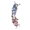
| Title | E.coli Hemolysin E (HlyE, ClyA, SheA) | ||||||
 Components Components | HEMOLYSIN E | ||||||
 Keywords Keywords | TOXIN / MEMBRANE PORE FORMER / CYTOLYSIN / HEMOLYSIN / PORES | ||||||
| Function / homology |  Function and homology information Function and homology informationmodulation of apoptotic process in another organism / hemolysis in another organism / toxin activity / periplasmic space / host cell plasma membrane / extracellular region / identical protein binding / membrane Similarity search - Function | ||||||
| Biological species |  | ||||||
| Method |  X-RAY DIFFRACTION / X-RAY DIFFRACTION /  SYNCHROTRON / SYNCHROTRON /  MIR / Resolution: 2 Å MIR / Resolution: 2 Å | ||||||
 Authors Authors | Wallace, A.J. / Stillman, T.J. / Atkins, A. / Jamieson, S.J. / Bullough, P.A. / Green, J. / Artymiuk, P.J. | ||||||
 Citation Citation |  Journal: Cell(Cambridge,Mass.) / Year: 2000 Journal: Cell(Cambridge,Mass.) / Year: 2000Title: E. Coli Hemolysin E (Hlye, Clya, Shea): X-Ray Crystal Structure of the Toxin and Observation of Membrane Pores by Electron Microscopy Authors: Wallace, A.J. / Stillman, T.J. / Atkins, A. / Jamieson, S.J. / Bullough, P.A. / Green, J. / Artymiuk, P.J. | ||||||
| History |
|
- Structure visualization
| Structure viewer | Molecule:  Molmil Molmil Jmol/JSmol Jmol/JSmol |
|---|
- Downloads & links
- Download
| PDBx/mmCIF format |  1qoy.cif.gz 1qoy.cif.gz | 79.4 KB | Display |  PDBx/mmCIF format PDBx/mmCIF format |
|---|---|---|---|---|
| PDB format |  pdb1qoy.ent.gz pdb1qoy.ent.gz | 59.5 KB | Display |  PDB format PDB format |
| PDBx/mmJSON format |  1qoy.json.gz 1qoy.json.gz | Tree view |  PDBx/mmJSON format PDBx/mmJSON format | |
| Others |  Other downloads Other downloads |
-Validation report
| Arichive directory |  https://data.pdbj.org/pub/pdb/validation_reports/qo/1qoy https://data.pdbj.org/pub/pdb/validation_reports/qo/1qoy ftp://data.pdbj.org/pub/pdb/validation_reports/qo/1qoy ftp://data.pdbj.org/pub/pdb/validation_reports/qo/1qoy | HTTPS FTP |
|---|
-Related structure data
| Similar structure data |
|---|
- Links
Links
- Assembly
| Deposited unit | 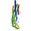
| ||||||||
|---|---|---|---|---|---|---|---|---|---|
| 1 |
| ||||||||
| Unit cell |
|
- Components
| #1: Protein | Mass: 35005.707 Da / Num. of mol.: 1 / Mutation: YES Source method: isolated from a genetically manipulated source Source: (gene. exp.)   |
|---|---|
| #2: Chemical | ChemComp-SO4 / |
| #3: Water | ChemComp-HOH / |
| Compound details | THE INTIAL 15 RESIDUES, GSPGISGGGGGILDS, ARE THE GST LINKER. THE T2A MUTATION WAS A RESULT OF A ...THE INTIAL 15 RESIDUES, GSPGISGGGG |
| Sequence details | THE SWISSPROT ENTRY HLYE_ECOLI REPORTS AN INCORRECT N-TERMINAL REGION OF THE PROTEIN THAT CONTAINS ...THE SWISSPROT ENTRY HLYE_ECOLI REPORTS AN INCORRECT N-TERMINAL REGION OF THE PROTEIN THAT CONTAINS A FRAMESHIFT |
-Experimental details
-Experiment
| Experiment | Method:  X-RAY DIFFRACTION / Number of used crystals: 1 X-RAY DIFFRACTION / Number of used crystals: 1 |
|---|
- Sample preparation
| Crystal | Density Matthews: 3.74 Å3/Da / Density % sol: 67.1 % | ||||||||||||||||||||||||||||||||||||||||||||||||
|---|---|---|---|---|---|---|---|---|---|---|---|---|---|---|---|---|---|---|---|---|---|---|---|---|---|---|---|---|---|---|---|---|---|---|---|---|---|---|---|---|---|---|---|---|---|---|---|---|---|
| Crystal grow | pH: 6 / Details: pH 6.00 | ||||||||||||||||||||||||||||||||||||||||||||||||
| Crystal grow | *PLUS pH: 6.8 / Method: vapor diffusion, hanging dropDetails: drop consists of equal volume of protein and reservoir solutions | ||||||||||||||||||||||||||||||||||||||||||||||||
| Components of the solutions | *PLUS
|
-Data collection
| Diffraction | Mean temperature: 100 K |
|---|---|
| Diffraction source | Source:  SYNCHROTRON / Site: SYNCHROTRON / Site:  SRS SRS  / Beamline: PX7.2 / Wavelength: 1.488 / Beamline: PX7.2 / Wavelength: 1.488 |
| Detector | Type: ADSC QUANTUM 4 CCD / Detector: CCD / Date: May 29, 1998 / Details: PLATINUM COATED MIRRORS |
| Radiation | Monochromator: GE(111) / Protocol: SINGLE WAVELENGTH / Monochromatic (M) / Laue (L): M / Scattering type: x-ray |
| Radiation wavelength | Wavelength: 1.488 Å / Relative weight: 1 |
| Reflection | Resolution: 2→17.8 Å / Num. obs: 31955 / % possible obs: 89.3 % / Redundancy: 2.5 % / Biso Wilson estimate: 22.5 Å2 / Rmerge(I) obs: 0.036 / Net I/σ(I): 12 |
| Reflection shell | Resolution: 2→2.05 Å / Redundancy: 2.1 % / Rmerge(I) obs: 0.056 / Mean I/σ(I) obs: 7.6 / % possible all: 72 |
| Reflection | *PLUS Num. measured all: 78696 |
| Reflection shell | *PLUS % possible obs: 72 % |
- Processing
| Software |
| ||||||||||||||||||||||||||||||||||||||||||||||||||||||||||||||||||||||||||||||||||||
|---|---|---|---|---|---|---|---|---|---|---|---|---|---|---|---|---|---|---|---|---|---|---|---|---|---|---|---|---|---|---|---|---|---|---|---|---|---|---|---|---|---|---|---|---|---|---|---|---|---|---|---|---|---|---|---|---|---|---|---|---|---|---|---|---|---|---|---|---|---|---|---|---|---|---|---|---|---|---|---|---|---|---|---|---|---|
| Refinement | Method to determine structure:  MIR / Resolution: 2→17.8 Å / SU ML: 0.095 / Cross valid method: THROUGHOUT / σ(F): 0 / ESU R: 0.158 / ESU R Free: 0.161 MIR / Resolution: 2→17.8 Å / SU ML: 0.095 / Cross valid method: THROUGHOUT / σ(F): 0 / ESU R: 0.158 / ESU R Free: 0.161
| ||||||||||||||||||||||||||||||||||||||||||||||||||||||||||||||||||||||||||||||||||||
| Displacement parameters | Biso mean: 29.7 Å2 | ||||||||||||||||||||||||||||||||||||||||||||||||||||||||||||||||||||||||||||||||||||
| Refinement step | Cycle: LAST / Resolution: 2→17.8 Å
| ||||||||||||||||||||||||||||||||||||||||||||||||||||||||||||||||||||||||||||||||||||
| Refine LS restraints |
| ||||||||||||||||||||||||||||||||||||||||||||||||||||||||||||||||||||||||||||||||||||
| Software | *PLUS Name: REFMAC / Classification: refinement | ||||||||||||||||||||||||||||||||||||||||||||||||||||||||||||||||||||||||||||||||||||
| Refine LS restraints | *PLUS
|
 Movie
Movie Controller





 PDBj
PDBj


