 Movie
Movie Controller


+ Open data

- Basic information
| Entry | Database: PDB / ID: 1qmf | ||||||
|---|---|---|---|---|---|---|---|
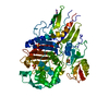
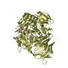
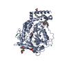
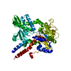
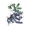
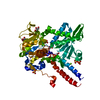
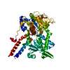
| Title | PENICILLIN-BINDING PROTEIN 2X (PBP-2X) ACYL-ENZYME COMPLEX | ||||||
 Components Components | PENICILLIN-BINDING PROTEIN 2X | ||||||
 Keywords Keywords | CELL CYCLE / PEPTIDOGLYCAN SYNTHESIS / CELL WALL | ||||||
| Function / homology |  Function and homology information Function and homology informationpenicillin binding / peptidoglycan biosynthetic process / cell wall organization / regulation of cell shape / cell division / response to antibiotic / plasma membrane Similarity search - Function | ||||||
| Biological species |   STREPTOCOCCUS PNEUMONIAE (bacteria) STREPTOCOCCUS PNEUMONIAE (bacteria) | ||||||
| Method |  X-RAY DIFFRACTION / SYNCHROTRON / MOLECULAR REPLACEMENT / Resolution: 2.8 Å X-RAY DIFFRACTION / SYNCHROTRON / MOLECULAR REPLACEMENT / Resolution: 2.8 Å | ||||||
 Authors Authors | Gordon, E.J. / Mouz, N. / Duee, E. / Dideberg, O. | ||||||
 Citation Citation | Journal: J.Mol.Biol. / Year: 2000 Title: The Crystal Structure of the Penicillin Binding Protein 2X from Streptococcus Pneumoniae and its Acyl-Enzyme Form: Implication in Drug Resistance Authors: Gordon, E.J. / Mouz, N. / Duee, E. / Dideberg, O. #1: Journal: Nat.Struct.Biol. / Year: 1996Title: X-Ray Structure of Streptococcus Pneumoniae Pbp2X, a Primary Penicillin Target Enzyme Authors: Pares, S. / Mouz, N. / Petillot, Y. / Hakenbeck, R. / Dideberg, O. #2: Journal: J.Mol.Biol. / Year: 1993 Title: Crystallization of a Genetically Engineered Water-Soluble Primary Penicillin Target Enzyme. The High Molecular Mass Pbp2X of Streptococcus Pneumoniae Authors: Charlier, P. / Buisson, G. / Dideberg, O. / Wierenga, J. / Keck, W. / Laible, G. / Hakenbeck, R. | ||||||
| History |
|
- Structure visualization
| Structure viewer | Molecule: MolmilJmol/JSmol |
|---|
- Downloads & links
-Download
| PDBx/mmCIF format | 1qmf.cif.gz | 125.4 KB | Display | PDBx/mmCIF format |
|---|---|---|---|---|
| PDB format | pdb1qmf.ent.gz | 93.7 KB | Display | PDB format |
| PDBx/mmJSON format | 1qmf.json.gz | Tree view | PDBx/mmJSON format | |
| Others |  Other downloads Other downloads |
-Validation report
| Arichive directory | https://data.pdbj.org/pub/pdb/validation_reports/qm/1qmfftp://data.pdbj.org/pub/pdb/validation_reports/qm/1qmf | HTTPS FTP |
|---|
-Related structure data


-Links
 PDBj
PDBj- Assembly
| Deposited unit | 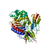
| ||||||||
|---|---|---|---|---|---|---|---|---|---|
| 1 |
| ||||||||
| Unit cell |
|
-Components
| #1: Protein | Mass: 76807.969 Da / Num. of mol.: 1 Source method: isolated from a genetically manipulated source Details: COMPLEXED WITH CEFUROXIME ANTIBIOTIC / Source: (gene. exp.) STREPTOCOCCUS PNEUMONIAE (bacteria) / Gene: PBP2X / Plasmid: PGEX / Production host: |
|---|---|
| #2: Chemical | ChemComp-CES /   Mass: 383.376 Da / Num. of mol.: 1 / Source method: obtained synthetically / Formula: C15H17N3O7S Mass: 383.376 Da / Num. of mol.: 1 / Source method: obtained synthetically / Formula: C15H17N3O7S |
| #3: Chemical | ChemComp-KEF /   Mass: 424.385 Da / Num. of mol.: 1 / Source method: obtained synthetically / Formula: C16H16N4O8S Mass: 424.385 Da / Num. of mol.: 1 / Source method: obtained synthetically / Formula: C16H16N4O8S |
| #4: Water | ChemComp-HOH /  Mass: 18.015 Da / Num. of mol.: 57 / Source method: isolated from a natural source / Formula: H2O Mass: 18.015 Da / Num. of mol.: 57 / Source method: isolated from a natural source / Formula: H2O |
| Has protein modification | Y |
-Experimental details
-Experiment
| Experiment | Method: X-RAY DIFFRACTION / Number of used crystals: 1 |
|---|
- Sample preparation
| Crystal | Density Matthews: 3.86 Å3/Da / Density % sol: 68 % | ||||||||||||||||||||||||||||||||||||
|---|---|---|---|---|---|---|---|---|---|---|---|---|---|---|---|---|---|---|---|---|---|---|---|---|---|---|---|---|---|---|---|---|---|---|---|---|---|
| Crystal grow | pH: 4.5 Details: 0.1M SODIUM ACETATE PH 4.5, 1.0-1.3M AMMONIUM SULFATE | ||||||||||||||||||||||||||||||||||||
| Crystal grow | *PLUS Temperature: 25 ℃ / pH: 8 / Method: vapor diffusion, hanging drop | ||||||||||||||||||||||||||||||||||||
| Components of the solutions | *PLUS
|
-Data collection
| Diffraction | Mean temperature: 100 K |
|---|---|
| Diffraction source | Source: SYNCHROTRON / Site: ESRF  / Beamline: BM14 / Wavelength: 0.98 / Beamline: BM14 / Wavelength: 0.98 |
| Detector | Type: MARRESEARCH / Detector: IMAGE PLATE / Date: Oct 15, 1995 |
| Radiation | Monochromator: SI(111) / Protocol: SINGLE WAVELENGTH / Monochromatic (M) / Laue (L): M / Scattering type: x-ray |
| Radiation wavelength | Wavelength: 0.98 Å / Relative weight: 1 |
| Reflection | Resolution: 2.8→50 Å / Num. obs: 29276 / % possible obs: 97.9 % / Observed criterion σ(I): 0 / Redundancy: 5.4 % / Biso Wilson estimate: 33 Å2 / Rmerge(I) obs: 0.092 / Rsym value: 0.092 |
| Reflection shell | Resolution: 2.8→2.95 Å / Rmerge(I) obs: 0.508 / % possible all: 92 |
| Reflection shell | *PLUS Highest resolution: 2.8 Å / % possible obs: 92.2 % |
- Processing
| Software |
| ||||||||||||||||||||||||||||||||||||||||||||||||||||||||||||||||||||||||||||||||
|---|---|---|---|---|---|---|---|---|---|---|---|---|---|---|---|---|---|---|---|---|---|---|---|---|---|---|---|---|---|---|---|---|---|---|---|---|---|---|---|---|---|---|---|---|---|---|---|---|---|---|---|---|---|---|---|---|---|---|---|---|---|---|---|---|---|---|---|---|---|---|---|---|---|---|---|---|---|---|---|---|---|
| Refinement | Method to determine structure: MOLECULAR REPLACEMENT Starting model: APO ENZYME Resolution: 2.8→50 Å / Rfactor Rfree error: 0.005 / Data cutoff high absF: 2313146.29 / Isotropic thermal model: RESTRAINED / Cross valid method: THROUGHOUT / σ(F): 0 / Stereochemistry target values: MLF Details: BULK SOLVENT MODEL USED. MEMBRANE ANCHOR HAS BEEN DELETED FROM CONSTRUCT. PROTEIN CRYSTALLISED CORRESPONDS TO RESIDUES 49-750. ALSO, RESIDUES 93-182, 233- 253, 621-631 ARE DISORDERED.
| ||||||||||||||||||||||||||||||||||||||||||||||||||||||||||||||||||||||||||||||||
| Solvent computation | Solvent model: FLAT MODEL / Bsol: 20.1677 Å2 / ksol: 0.317581 e/Å3 | ||||||||||||||||||||||||||||||||||||||||||||||||||||||||||||||||||||||||||||||||
| Displacement parameters | Biso mean: 52.4 Å2
| ||||||||||||||||||||||||||||||||||||||||||||||||||||||||||||||||||||||||||||||||
| Refine analyze |
| ||||||||||||||||||||||||||||||||||||||||||||||||||||||||||||||||||||||||||||||||
| Refinement step | Cycle: LAST / Resolution: 2.8→50 Å
| ||||||||||||||||||||||||||||||||||||||||||||||||||||||||||||||||||||||||||||||||
| Refine LS restraints |
| ||||||||||||||||||||||||||||||||||||||||||||||||||||||||||||||||||||||||||||||||
| LS refinement shell | Resolution: 2.8→2.98 Å / Rfactor Rfree error: 0.018 / Total num. of bins used: 6
| ||||||||||||||||||||||||||||||||||||||||||||||||||||||||||||||||||||||||||||||||
| Xplor file |
| ||||||||||||||||||||||||||||||||||||||||||||||||||||||||||||||||||||||||||||||||
| Software | *PLUS Name: CNS / Version: 0.5 / Classification: refinement | ||||||||||||||||||||||||||||||||||||||||||||||||||||||||||||||||||||||||||||||||
| Refine LS restraints | *PLUS
| ||||||||||||||||||||||||||||||||||||||||||||||||||||||||||||||||||||||||||||||||
| LS refinement shell | *PLUS Rfactor Rfree: 0.38 / Rfactor obs: 0.341 |
