 Movie
Movie Controller

[English] 日本語
 Yorodumi
- PDB-6s48: AvaII RESTRICTION ENDONUCLEASE IN COMPLEX WITH PARTIALLY CLEAVED dsDNA -
+ Open data

- Basic information
| Entry | Database: PDB / ID: 6s48 | ||||||
|---|---|---|---|---|---|---|---|
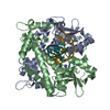
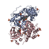
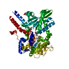
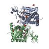
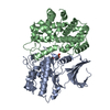
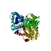
| Title | AvaII RESTRICTION ENDONUCLEASE IN COMPLEX WITH PARTIALLY CLEAVED dsDNA | ||||||
 Components Components |
| ||||||
 Keywords Keywords | HYDROLASE / RESTRICTION ENDONUCLEASE / DNA COMPLEX / A-DNA / PD-(D/E)XK | ||||||
| Function / homology |  Function and homology information Function and homology informationtype II site-specific deoxyribonuclease activity / DNA restriction-modification system / DNA binding / metal ion binding Similarity search - Function | ||||||
| Biological species |  Nostoc sp. PCC 7120 = FACHB-418 (bacteria) Nostoc sp. PCC 7120 = FACHB-418 (bacteria)synthetic construct (others) | ||||||
| Method |  X-RAY DIFFRACTION / SYNCHROTRON / MOLECULAR REPLACEMENT / Resolution: 1.9 Å X-RAY DIFFRACTION / SYNCHROTRON / MOLECULAR REPLACEMENT / Resolution: 1.9 Å | ||||||
 Authors Authors | Kisiala, M. / Kowalska, M. / Korza, H. / Czapinska, H. / Bochtler, M. | ||||||
 Citation Citation | Journal: Nucleic Acids Res. / Year: 2020 Title: Restriction endonucleases that cleave RNA/DNA heteroduplexes bind dsDNA in A-like conformation. Authors: Kisiala, M. / Kowalska, M. / Pastor, M. / Korza, H.J. / Czapinska, H. / Bochtler, M. #1: Journal: Nucleic Acids Res. / Year: 2010 Title: Sequence-Specific Cleavage Of Rna By Type Ii Restriction Enzymes. Authors: Murray, I.A. / Stickel, S.K. / Roberts, R.J. #2: Journal: Nucleic Acids Res. / Year: 1980 Title: Cleavage of DNA.RNA hybrids by type II restriction enzymes. Authors: Molloy, P.L. / Symons, R.H. #3: Journal: J. Biol. Chem. / Year: 2005Title: Crystal Structures Of Type Ii Restriction Endonuclease Ecoo109I And Its Complex With Cognate Dna. Authors: Hashimoto, H. / Shimizu, T. / Imasaki, T. / Kato, M. / Shichijo, N. / Kita, K. / Sato, M. #4: Journal: Biophys. J. / Year: 2009 Title: Intrinsic dynamics of restriction endonuclease EcoO109I studied by molecular dynamics simulations and X-ray scattering data analysis. Authors: Oroguchi, T. / Hashimoto, H. / Shimizu, T. / Sato, M. / Ikeguchi, M. | ||||||
| History |
|
- Structure visualization
| Structure viewer | Molecule: MolmilJmol/JSmol |
|---|
- Downloads & links
-Download
| PDBx/mmCIF format | 6s48.cif.gz | 153.6 KB | Display | PDBx/mmCIF format |
|---|---|---|---|---|
| PDB format | pdb6s48.ent.gz | 115.3 KB | Display | PDB format |
| PDBx/mmJSON format | 6s48.json.gz | Tree view | PDBx/mmJSON format | |
| Others |  Other downloads Other downloads |
-Validation report
| Arichive directory | https://data.pdbj.org/pub/pdb/validation_reports/s4/6s48ftp://data.pdbj.org/pub/pdb/validation_reports/s4/6s48 | HTTPS FTP |
|---|
-Related structure data
| Related structure data | 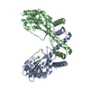 6s58C 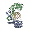 6g3bS S: Starting model for refinement C: citing same article ( |
|---|---|
| Similar structure data |


-Links
 PDBj
PDBj


- Assembly
| Deposited unit | 
| ||||||||
|---|---|---|---|---|---|---|---|---|---|
| 1 |
| ||||||||
| Unit cell |
|
-Components
-Protein , 1 types, 2 molecules AB
| #1: Protein | Mass: 27161.797 Da / Num. of mol.: 2 Source method: isolated from a genetically manipulated source Source: (gene. exp.) Nostoc sp. PCC 7120 = FACHB-418 (bacteria)Gene: alr0933 / Plasmid: pET28a / Production host: |
|---|
-DNA chain , 5 types, 7 molecules CDHEIFG
| #2: DNA chain | Mass: 3349.197 Da / Num. of mol.: 1 / Source method: obtained synthetically / Source: (synth.) synthetic construct (others) | ||||||
|---|---|---|---|---|---|---|---|
| #3: DNA chain | Mass: 1230.854 Da / Num. of mol.: 2 / Source method: obtained synthetically / Source: (synth.) synthetic construct (others) #4: DNA chain | Mass: 2082.400 Da / Num. of mol.: 2 / Source method: obtained synthetically / Source: (synth.) synthetic construct (others) #5: DNA chain | | Mass: 1230.854 Da / Num. of mol.: 1 / Source method: obtained synthetically / Source: (synth.) synthetic construct (others) #6: DNA chain | | Mass: 2073.386 Da / Num. of mol.: 1 / Source method: obtained synthetically / Source: (synth.) synthetic construct (others) |
-Non-polymers , 6 types, 497 molecules 


| #7: Chemical | ChemComp-CA /  Mass: 40.078 Da / Num. of mol.: 5 / Source method: obtained synthetically / Formula: Ca Mass: 40.078 Da / Num. of mol.: 5 / Source method: obtained synthetically / Formula: Ca#8: Chemical |  Mass: 78.133 Da / Num. of mol.: 2 / Source method: isolated from a natural source / Formula: C2H6OS Mass: 78.133 Da / Num. of mol.: 2 / Source method: isolated from a natural source / Formula: C2H6OS#9: Chemical | ChemComp-1PE / |  Mass: 238.278 Da / Num. of mol.: 1 / Source method: obtained synthetically / Formula: C10H22O6 / Comment: precipitant*YM Mass: 238.278 Da / Num. of mol.: 1 / Source method: obtained synthetically / Formula: C10H22O6 / Comment: precipitant*YM#10: Chemical | ChemComp-PGE / |  Mass: 150.173 Da / Num. of mol.: 1 / Source method: obtained synthetically / Formula: C6H14O4 Mass: 150.173 Da / Num. of mol.: 1 / Source method: obtained synthetically / Formula: C6H14O4#11: Chemical | ChemComp-SER / |  Type: L-peptide linking / Mass: 105.093 Da / Num. of mol.: 1 / Source method: isolated from a natural source / Formula: C3H7NO3 Type: L-peptide linking / Mass: 105.093 Da / Num. of mol.: 1 / Source method: isolated from a natural source / Formula: C3H7NO3#12: Water | ChemComp-HOH / | Mass: 18.015 Da / Num. of mol.: 487 / Source method: isolated from a natural source / Formula: H2O |
|---|
-Details
| Has ligand of interest | N |
|---|
-Experimental details
-Experiment
| Experiment | Method: X-RAY DIFFRACTION / Number of used crystals: 1 |
|---|
- Sample preparation
| Crystal grow | Temperature: 291 K / Method: vapor diffusion, sitting drop / pH: 6.5 Details: Morpheus screen H1 conditions: 0.02 M of L-Na-glutamate, alanine (racemic), glycine, lysine HCl (racemic), serine (racemic), 0.1 M buffer (1 M Imidazole, 1 M MES, pH 6.5), 30% precipitant ...Details: Morpheus screen H1 conditions: 0.02 M of L-Na-glutamate, alanine (racemic), glycine, lysine HCl (racemic), serine (racemic), 0.1 M buffer (1 M Imidazole, 1 M MES, pH 6.5), 30% precipitant (20% w/v PEG 20 000, 40% v/v PEG MME 550) |
|---|
-Data collection
| Diffraction | Mean temperature: 100 K / Serial crystal experiment: N |
|---|---|
| Diffraction source | Source: SYNCHROTRON / Site: BESSY  / Beamline: 14.1 / Wavelength: 0.9184 Å / Beamline: 14.1 / Wavelength: 0.9184 Å |
| Detector | Type: DECTRIS PILATUS 6M / Detector: PIXEL / Date: May 1, 2019 |
| Radiation | Protocol: SINGLE WAVELENGTH / Monochromatic (M) / Laue (L): M / Scattering type: x-ray |
| Radiation wavelength | Wavelength: 0.9184 Å / Relative weight: 1 |
| Reflection | Resolution: 1.9→40.07 Å / Num. obs: 36585 / % possible obs: 98.5 % / Redundancy: 3.8 % / Biso Wilson estimate: 36.1 Å2 / CC1/2: 0.998 / Rrim(I) all: 0.094 / Rsym value: 0.081 / Net I/σ(I): 10.55 |
| Reflection shell | Resolution: 1.9→2.01 Å / Redundancy: 3.7 % / Mean I/σ(I) obs: 1.43 / Num. unique obs: 5824 / CC1/2: 0.615 / Rrim(I) all: 0.943 / Rsym value: 0.807 / % possible all: 97.6 |
- Processing
| Software |
| ||||||||||||||||||||||||||||||||||||||||||||||||||||||||||||||||||||||||||||||||||||||||||||||||||||||||||||||||||||||||||||||||||||||||||||||||||||||||||||||||||||||||||||||||||||||
|---|---|---|---|---|---|---|---|---|---|---|---|---|---|---|---|---|---|---|---|---|---|---|---|---|---|---|---|---|---|---|---|---|---|---|---|---|---|---|---|---|---|---|---|---|---|---|---|---|---|---|---|---|---|---|---|---|---|---|---|---|---|---|---|---|---|---|---|---|---|---|---|---|---|---|---|---|---|---|---|---|---|---|---|---|---|---|---|---|---|---|---|---|---|---|---|---|---|---|---|---|---|---|---|---|---|---|---|---|---|---|---|---|---|---|---|---|---|---|---|---|---|---|---|---|---|---|---|---|---|---|---|---|---|---|---|---|---|---|---|---|---|---|---|---|---|---|---|---|---|---|---|---|---|---|---|---|---|---|---|---|---|---|---|---|---|---|---|---|---|---|---|---|---|---|---|---|---|---|---|---|---|---|---|
| Refinement | Method to determine structure: MOLECULAR REPLACEMENT Starting model: 6G3B Resolution: 1.9→40.07 Å / Cor.coef. Fo:Fc: 0.968 / Cor.coef. Fo:Fc free: 0.952 / SU B: 5.795 / SU ML: 0.157 / Cross valid method: THROUGHOUT / ESU R: 0.22 / ESU R Free: 0.166 Details: HYDROGEN ATOMS HAVE BEEN ADDED IN THE RIDING POSITIONS. THE ION IDENTITY WAS ASSIGNED TENTATIVELY. THE RESOLUTION OF THE STRUCTURE WAS NOT GOOD ENOUGH FOR ITS EXPERIMENTAL VALIDATION. THE ...Details: HYDROGEN ATOMS HAVE BEEN ADDED IN THE RIDING POSITIONS. THE ION IDENTITY WAS ASSIGNED TENTATIVELY. THE RESOLUTION OF THE STRUCTURE WAS NOT GOOD ENOUGH FOR ITS EXPERIMENTAL VALIDATION. THE ION IDENTIFICATION THROUGH LONG WAVELENGTH DATA COLLECTION WAS ATTEMPTED BUT THE ANOMALOUS SIGNAL WAS VERY WEAK. THE CYSTEINE MODIFICATION WAS MODELED AS 2-MERCAPTOETHANOL, BUT MAY AS WELL CORRESPOND TO DITHIOTHREITOL. BOTH AGENTS WERE PRESENT AT CERTAIN PURIFICATION STAGES. FREE SERINE FROM THE CRYSTALLIZATION BUFFER WAS MODELED IN THE DENSITY BUT ITS IDENTITY IS VERY UNCERTAIN.
| ||||||||||||||||||||||||||||||||||||||||||||||||||||||||||||||||||||||||||||||||||||||||||||||||||||||||||||||||||||||||||||||||||||||||||||||||||||||||||||||||||||||||||||||||||||||
| Solvent computation | Ion probe radii: 0.8 Å / Shrinkage radii: 0.8 Å / VDW probe radii: 1.2 Å | ||||||||||||||||||||||||||||||||||||||||||||||||||||||||||||||||||||||||||||||||||||||||||||||||||||||||||||||||||||||||||||||||||||||||||||||||||||||||||||||||||||||||||||||||||||||
| Displacement parameters | Biso mean: 35.854 Å2
| ||||||||||||||||||||||||||||||||||||||||||||||||||||||||||||||||||||||||||||||||||||||||||||||||||||||||||||||||||||||||||||||||||||||||||||||||||||||||||||||||||||||||||||||||||||||
| Refinement step | Cycle: LAST / Resolution: 1.9→40.07 Å
| ||||||||||||||||||||||||||||||||||||||||||||||||||||||||||||||||||||||||||||||||||||||||||||||||||||||||||||||||||||||||||||||||||||||||||||||||||||||||||||||||||||||||||||||||||||||
| Refine LS restraints |
|