 Movie
Movie Controller


+ Open data

- Basic information
| Entry | Database: PDB / ID: 1qhw | |||||||||
|---|---|---|---|---|---|---|---|---|---|---|
| Title | PURPLE ACID PHOSPHATASE FROM RAT BONE | |||||||||
 Components Components | PROTEIN (PURPLE ACID PHOSPHATASE) | |||||||||
 Keywords Keywords | HYDROLASE / METAL PHOSPHATASE | |||||||||
| Function / homology |  Function and homology information Function and homology informationnegative regulation of superoxide anion generation / Vitamin B2 (riboflavin) metabolism / cellular response to zinc ion starvation / response to macrophage colony-stimulating factor / acid phosphatase / acid phosphatase activity / response to L-ascorbic acid / negative regulation of interleukin-12 production / negative regulation of cell adhesion / negative regulation of nitric oxide biosynthetic process ...negative regulation of superoxide anion generation / Vitamin B2 (riboflavin) metabolism / cellular response to zinc ion starvation / response to macrophage colony-stimulating factor / acid phosphatase / acid phosphatase activity / response to L-ascorbic acid / negative regulation of interleukin-12 production / negative regulation of cell adhesion / negative regulation of nitric oxide biosynthetic process / response to cholesterol / bone morphogenesis / dephosphorylation / response to zinc ion / negative regulation of interleukin-1 beta production / bone resorption / negative regulation of tumor necrosis factor production / multicellular organismal response to stress / response to mechanical stimulus / osteoclast differentiation / response to cytokine / ferric iron binding / ossification / ferrous iron binding / response to insulin / negative regulation of inflammatory response / response to lipopolysaccharide / response to ethanol / lysosome / defense response to Gram-positive bacterium / positive regulation of cell migration / hydrolase activity / : Similarity search - Function | |||||||||
| Biological species |  | |||||||||
| Method |  X-RAY DIFFRACTION / SYNCHROTRON / MOLECULAR REPLACEMENT / Resolution: 2.2 Å X-RAY DIFFRACTION / SYNCHROTRON / MOLECULAR REPLACEMENT / Resolution: 2.2 Å | |||||||||
 Authors Authors | Lindqvist, Y. / Johansson, E. / Kaija, H. / Vihko, P. / Schneider, G. | |||||||||
 Citation Citation | Journal: J.Mol.Biol. / Year: 1999 Title: Three-dimensional structure of a mammalian purple acid phosphatase at 2.2 A resolution with a mu-(hydr)oxo bridged di-iron center. Authors: Lindqvist, Y. / Johansson, E. / Kaija, H. / Vihko, P. / Schneider, G. #1: Journal: J.Bone Miner.Res. / Year: 1999Title: Tartrate-Resistant Bone Acid Phosphatase: Large-Scale Production and Purification of the Recombinant Enzyme, Characterization, and Crystallization Authors: Kaija, H. / Jia, J. / Lindqvist, Y. / Andersson, G. / Vihko, P. | |||||||||
| History |
|
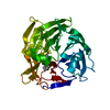
- Structure visualization
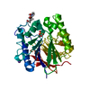
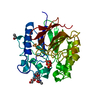
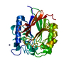
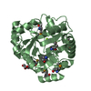
| Structure viewer | Molecule: MolmilJmol/JSmol |
|---|
- Downloads & links
-Download
| PDBx/mmCIF format | 1qhw.cif.gz | 77.5 KB | Display | PDBx/mmCIF format |
|---|---|---|---|---|
| PDB format | pdb1qhw.ent.gz | 56.3 KB | Display | PDB format |
| PDBx/mmJSON format | 1qhw.json.gz | Tree view | PDBx/mmJSON format | |
| Others |  Other downloads Other downloads |
-Validation report
| Arichive directory | https://data.pdbj.org/pub/pdb/validation_reports/qh/1qhwftp://data.pdbj.org/pub/pdb/validation_reports/qh/1qhw | HTTPS FTP |
|---|
-Related structure data
| Related structure data |  4kbpS S: Starting model for refinement |
|---|---|
| Similar structure data |


-Links
 PDBj
PDBj
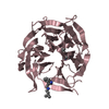
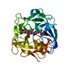
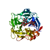
- Assembly
| Deposited unit | 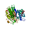
| ||||||||
|---|---|---|---|---|---|---|---|---|---|
| 1 |
| ||||||||
| Unit cell |
|
-Components
-Protein / Sugars , 2 types, 2 molecules A
| #1: Protein | Mass: 36769.984 Da / Num. of mol.: 1 Source method: isolated from a genetically manipulated source Details: GLYCOSIDIC LINK BETWEEN ASN 118 AND NAG 344 GLYCOSIDIC LINK BETWEEN NAG 344 AND NAG 345 Source: (gene. exp.)   Spodoptera frugiperda (fall armyworm) / Strain (production host): SF9 / References: UniProt: P29288, acid phosphatase Spodoptera frugiperda (fall armyworm) / Strain (production host): SF9 / References: UniProt: P29288, acid phosphatase |
|---|---|
| #2: Polysaccharide | 2-acetamido-2-deoxy-beta-D-glucopyranose-(1-4)-2-acetamido-2-deoxy-beta-D-glucopyranose Source method: isolated from a genetically manipulated source |
-Non-polymers , 4 types, 109 molecules 


| #3: Chemical |  Mass: 55.845 Da / Num. of mol.: 2 / Source method: obtained synthetically / Formula: Fe Mass: 55.845 Da / Num. of mol.: 2 / Source method: obtained synthetically / Formula: Fe#4: Chemical |  Mass: 65.409 Da / Num. of mol.: 2 / Source method: obtained synthetically / Formula: Zn Mass: 65.409 Da / Num. of mol.: 2 / Source method: obtained synthetically / Formula: Zn#5: Chemical |  Mass: 96.063 Da / Num. of mol.: 2 / Source method: obtained synthetically / Formula: SO4 Mass: 96.063 Da / Num. of mol.: 2 / Source method: obtained synthetically / Formula: SO4#6: Water | ChemComp-HOH / | Mass: 18.015 Da / Num. of mol.: 103 / Source method: isolated from a natural source / Formula: H2O |
|---|
-Details
| Has protein modification | Y |
|---|
-Experimental details
-Experiment
| Experiment | Method: X-RAY DIFFRACTION / Number of used crystals: 2 |
|---|
- Sample preparation
| Crystal | Density Matthews: 2.55 Å3/Da / Density % sol: 45 % | ||||||||||||||||||||||||||||||
|---|---|---|---|---|---|---|---|---|---|---|---|---|---|---|---|---|---|---|---|---|---|---|---|---|---|---|---|---|---|---|---|
| Crystal grow | pH: 7.5 / Details: pH 7.5 | ||||||||||||||||||||||||||||||
| Crystal | *PLUS | ||||||||||||||||||||||||||||||
| Crystal grow | *PLUS Method: vapor diffusion, hanging drop / Details: Kaija, H., (1999) J.Bone Miner.Res., 14, 424. | ||||||||||||||||||||||||||||||
| Components of the solutions | *PLUS
|
-Data collection
| Diffraction | Mean temperature: 100 K |
|---|---|
| Diffraction source | Source: SYNCHROTRON / Site: ESRF  / Beamline: ID13 / Wavelength: 0.7816 / Beamline: ID13 / Wavelength: 0.7816 |
| Detector | Detector: CCD |
| Radiation | Protocol: SINGLE WAVELENGTH / Monochromatic (M) / Laue (L): M / Scattering type: x-ray |
| Radiation wavelength | Wavelength: 0.7816 Å / Relative weight: 1 |
| Reflection | Resolution: 2.2→40 Å / Num. obs: 17244 / % possible obs: 93.3 % / Observed criterion σ(I): 0 / Redundancy: 3.7 % / Biso Wilson estimate: 31 Å2 / Rmerge(I) obs: 0.127 / Net I/σ(I): 6.2 |
| Reflection shell | Resolution: 2.2→2.3 Å / Rmerge(I) obs: 0.344 / Mean I/σ(I) obs: 3.2 / % possible all: 84.4 |
| Reflection | *PLUS Num. measured all: 63605 |
| Reflection shell | *PLUS % possible obs: 84.4 % |
- Processing
| Software |
| ||||||||||||||||||||||||||||||||||||||||||||||||||||||||||||||||||||||||||||||||
|---|---|---|---|---|---|---|---|---|---|---|---|---|---|---|---|---|---|---|---|---|---|---|---|---|---|---|---|---|---|---|---|---|---|---|---|---|---|---|---|---|---|---|---|---|---|---|---|---|---|---|---|---|---|---|---|---|---|---|---|---|---|---|---|---|---|---|---|---|---|---|---|---|---|---|---|---|---|---|---|---|---|
| Refinement | Method to determine structure: MOLECULAR REPLACEMENT Starting model: BASED ON PDB ENTRY 4KBP Resolution: 2.2→40 Å / Rfactor Rfree error: 0.009 / Data cutoff high rms absF: 1502012.17 / Isotropic thermal model: RESTRAINED / Cross valid method: THROUGHOUT / σ(F): 0
| ||||||||||||||||||||||||||||||||||||||||||||||||||||||||||||||||||||||||||||||||
| Solvent computation | Solvent model: FLAT MODEL / Bsol: 47 Å2 / ksol: 0.37 e/Å3 | ||||||||||||||||||||||||||||||||||||||||||||||||||||||||||||||||||||||||||||||||
| Displacement parameters | Biso mean: 35.3 Å2
| ||||||||||||||||||||||||||||||||||||||||||||||||||||||||||||||||||||||||||||||||
| Refine analyze |
| ||||||||||||||||||||||||||||||||||||||||||||||||||||||||||||||||||||||||||||||||
| Refinement step | Cycle: LAST / Resolution: 2.2→40 Å
| ||||||||||||||||||||||||||||||||||||||||||||||||||||||||||||||||||||||||||||||||
| Refine LS restraints |
| ||||||||||||||||||||||||||||||||||||||||||||||||||||||||||||||||||||||||||||||||
| LS refinement shell | Resolution: 2.2→2.34 Å / Rfactor Rfree error: 0.026 / Total num. of bins used: 6
| ||||||||||||||||||||||||||||||||||||||||||||||||||||||||||||||||||||||||||||||||
| Xplor file |
| ||||||||||||||||||||||||||||||||||||||||||||||||||||||||||||||||||||||||||||||||
| Software | *PLUS Name: CNS / Version: 0.5 / Classification: refinement | ||||||||||||||||||||||||||||||||||||||||||||||||||||||||||||||||||||||||||||||||
| Refinement | *PLUS Highest resolution: 2.2 Å / Lowest resolution: 40 Å / σ(F): 0 / % reflection Rfree: 5.1 % | ||||||||||||||||||||||||||||||||||||||||||||||||||||||||||||||||||||||||||||||||
| Solvent computation | *PLUS | ||||||||||||||||||||||||||||||||||||||||||||||||||||||||||||||||||||||||||||||||
| Displacement parameters | *PLUS Biso mean: 35.3 Å2 | ||||||||||||||||||||||||||||||||||||||||||||||||||||||||||||||||||||||||||||||||
| Refine LS restraints | *PLUS
| ||||||||||||||||||||||||||||||||||||||||||||||||||||||||||||||||||||||||||||||||
| LS refinement shell | *PLUS Highest resolution: 2.2 Å / Rfactor Rfree: 0.317 / % reflection Rfree: 5.9 % / Rfactor Rwork: 0.264 / Rfactor obs: 0.264 |
