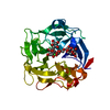
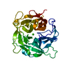
 Movie
Movie Controller


+ Open data

- Basic information
| Entry | Database: PDB / ID: 1ute | |||||||||
|---|---|---|---|---|---|---|---|---|---|---|
| Title | PIG PURPLE ACID PHOSPHATASE COMPLEXED WITH PHOSPHATE | |||||||||
 Components Components | PROTEIN (II PURPLE ACID PHOSPHATASE) | |||||||||
 Keywords Keywords | HYDROLASE / PURPLE ACID PHOSPHATASE / TARTRATE RESISTANT ACID PHOSPHATASE / METALLOENZYME / UTEROFERRIN | |||||||||
| Function / homology |  Function and homology information Function and homology informationVitamin B2 (riboflavin) metabolism / acid phosphatase / acid phosphatase activity / ferric iron binding / iron ion transport / ferrous iron binding / extracellular region Similarity search - Function | |||||||||
| Biological species |  | |||||||||
| Method |  X-RAY DIFFRACTION / MIR / Resolution: 1.55 Å X-RAY DIFFRACTION / MIR / Resolution: 1.55 Å | |||||||||
 Authors Authors | Guddat, L.W. / Mcalpine, A. / Hume, D. / Hamilton, S. / De Jersey, J. / Martin, J.L. | |||||||||
 Citation Citation | Journal: Structure Fold.Des. / Year: 1999 Title: Crystal structure of mammalian purple acid phosphatase. Authors: Guddat, L.W. / McAlpine, A.S. / Hume, D. / Hamilton, S. / de Jersey, J. / Martin, J.L. #1: Journal: Science / Year: 1995Title: Crystal Structure of a Purple Acid Phosphatase Containing a Dinuclear Fe(III)- Zn(II) Active Site Authors: Strater, N. / Klabunde, T. / Tucker, P. / Witzel, H. / Krebs, B. | |||||||||
| History |
|
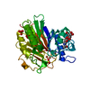
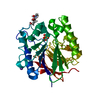
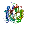
- Structure visualization
| Structure viewer | Molecule: MolmilJmol/JSmol |
|---|
- Downloads & links
-Download
| PDBx/mmCIF format | 1ute.cif.gz | 82.7 KB | Display | PDBx/mmCIF format |
|---|---|---|---|---|
| PDB format | pdb1ute.ent.gz | 60.3 KB | Display | PDB format |
| PDBx/mmJSON format | 1ute.json.gz | Tree view | PDBx/mmJSON format | |
| Others |  Other downloads Other downloads |
-Validation report
| Arichive directory | https://data.pdbj.org/pub/pdb/validation_reports/ut/1uteftp://data.pdbj.org/pub/pdb/validation_reports/ut/1ute | HTTPS FTP |
|---|
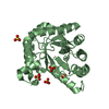
-Related structure data
| Similar structure data |
|---|


-Links
 PDBj
PDBj

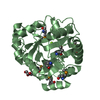
- Assembly
| Deposited unit | 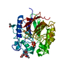
| ||||||||
|---|---|---|---|---|---|---|---|---|---|
| 1 |
| ||||||||
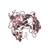
| Unit cell |
|
-Components
-Protein / Sugars , 2 types, 2 molecules A
| #1: Protein | Mass: 35154.066 Da / Num. of mol.: 1 / Source method: isolated from a natural source Details: PO4 PHOSPHATE FE+3 BINUCLEAR METAL CENTRE OXO OXYGEN BRIDGE BETWEEN TWO FE+3. IPA ISOPROPANOL Source: (natural) |
|---|---|
| #2: Polysaccharide | 2-acetamido-2-deoxy-beta-D-glucopyranose-(1-4)-2-acetamido-2-deoxy-beta-D-glucopyranose Source method: isolated from a genetically manipulated source |
-Non-polymers , 4 types, 342 molecules 


| #3: Chemical | ChemComp-PO4 /  Mass: 94.971 Da / Num. of mol.: 1 / Source method: obtained synthetically / Formula: PO4 Mass: 94.971 Da / Num. of mol.: 1 / Source method: obtained synthetically / Formula: PO4 |
|---|---|
| #4: Chemical | ChemComp-FEO /  Mass: 127.689 Da / Num. of mol.: 1 / Source method: obtained synthetically / Formula: Fe2O Mass: 127.689 Da / Num. of mol.: 1 / Source method: obtained synthetically / Formula: Fe2O |
| #5: Chemical | ChemComp-IPA /  Mass: 60.095 Da / Num. of mol.: 1 / Source method: obtained synthetically / Formula: C3H8O Mass: 60.095 Da / Num. of mol.: 1 / Source method: obtained synthetically / Formula: C3H8O |
| #6: Water | ChemComp-HOH / Mass: 18.015 Da / Num. of mol.: 339 / Source method: isolated from a natural source / Formula: H2O |
-Details
| Has protein modification | Y |
|---|
-Experimental details
-Experiment
| Experiment | Method: X-RAY DIFFRACTION / Number of used crystals: 1 |
|---|
- Sample preparation
| Crystal | Density Matthews: 2.3 Å3/Da / Density % sol: 53 % | ||||||||||||||||||||||||||||||
|---|---|---|---|---|---|---|---|---|---|---|---|---|---|---|---|---|---|---|---|---|---|---|---|---|---|---|---|---|---|---|---|
| Crystal grow | pH: 5 Details: 25% (W/V) PEG3350, 0.1 M LICL, 5% (V/V)ISOPROPANOL, 0.1 M SODIUM CITRATE PH 5.0. PROTEIN CONCENTRATION 38 MG/ML. PROTEIN CONCENTRATION 38MG/ML. | ||||||||||||||||||||||||||||||
| Crystal | *PLUS | ||||||||||||||||||||||||||||||
| Crystal grow | *PLUS Method: vapor diffusion, hanging drop | ||||||||||||||||||||||||||||||
| Components of the solutions | *PLUS
|
-Data collection
| Diffraction | Mean temperature: 100 K |
|---|---|
| Diffraction source | Source: ROTATING ANODE / Type: RIGAKU RU200 / Wavelength: 1.5418 |
| Detector | Type: RIGAKU RAXIS / Detector: IMAGE PLATE / Date: Jul 15, 1998 / Details: MIRRORS |
| Radiation | Monochromator: NI FILTER / Protocol: SINGLE WAVELENGTH / Monochromatic (M) / Laue (L): M / Scattering type: x-ray |
| Radiation wavelength | Wavelength: 1.5418 Å / Relative weight: 1 |
| Reflection | Resolution: 1.55→100 Å / Num. obs: 140370 / % possible obs: 89.4 % / Observed criterion σ(I): 0 / Redundancy: 3 % / Biso Wilson estimate: 15 Å2 / Rmerge(I) obs: 0.059 / Net I/σ(I): 9.6 |
| Reflection shell | Resolution: 1.55→1.61 Å / Redundancy: 1.4 % / Rmerge(I) obs: 0.271 / Mean I/σ(I) obs: 2.1 / % possible all: 57 |
| Reflection | *PLUS Num. obs: 46161 / Num. measured all: 140370 |
| Reflection shell | *PLUS % possible obs: 57.4 % |
- Processing
| Software |
| ||||||||||||||||||||||||||||||||||||||||||||||||||||||||||||||||||||||||||||||||
|---|---|---|---|---|---|---|---|---|---|---|---|---|---|---|---|---|---|---|---|---|---|---|---|---|---|---|---|---|---|---|---|---|---|---|---|---|---|---|---|---|---|---|---|---|---|---|---|---|---|---|---|---|---|---|---|---|---|---|---|---|---|---|---|---|---|---|---|---|---|---|---|---|---|---|---|---|---|---|---|---|---|
| Refinement | Method to determine structure: MIR / Resolution: 1.55→100 Å / Data cutoff high absF: 100000 / Data cutoff low absF: 0.001 / Cross valid method: THROUGHOUT / σ(F): 1
| ||||||||||||||||||||||||||||||||||||||||||||||||||||||||||||||||||||||||||||||||
| Displacement parameters | Biso mean: 20 Å2 | ||||||||||||||||||||||||||||||||||||||||||||||||||||||||||||||||||||||||||||||||
| Refinement step | Cycle: LAST / Resolution: 1.55→100 Å
| ||||||||||||||||||||||||||||||||||||||||||||||||||||||||||||||||||||||||||||||||
| Refine LS restraints |
| ||||||||||||||||||||||||||||||||||||||||||||||||||||||||||||||||||||||||||||||||
| LS refinement shell | Resolution: 1.55→1.62 Å / Total num. of bins used: 8
| ||||||||||||||||||||||||||||||||||||||||||||||||||||||||||||||||||||||||||||||||
| Xplor file |
| ||||||||||||||||||||||||||||||||||||||||||||||||||||||||||||||||||||||||||||||||
| Software | *PLUS Name: X-PLOR / Version: 3.851 / Classification: refinement | ||||||||||||||||||||||||||||||||||||||||||||||||||||||||||||||||||||||||||||||||
| Refinement | *PLUS σ(F): 1 / % reflection Rfree: 10 % | ||||||||||||||||||||||||||||||||||||||||||||||||||||||||||||||||||||||||||||||||
| Solvent computation | *PLUS | ||||||||||||||||||||||||||||||||||||||||||||||||||||||||||||||||||||||||||||||||
| Displacement parameters | *PLUS Biso mean: 20 Å2 | ||||||||||||||||||||||||||||||||||||||||||||||||||||||||||||||||||||||||||||||||
| Refine LS restraints | *PLUS
| ||||||||||||||||||||||||||||||||||||||||||||||||||||||||||||||||||||||||||||||||
| LS refinement shell | *PLUS Rfactor Rfree: 0.354 / % reflection Rfree: 9.4 % / Rfactor Rwork: 0.324 |
