 Movie
Movie Controller

[English] 日本語
 Yorodumi
- PDB-6s5j: Strictosidine Synthase from Ophiorrhiza pumila in complex with (S... -
+ Open data

- Basic information
| Entry | Database: PDB / ID: 6s5j | ||||||
|---|---|---|---|---|---|---|---|
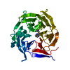
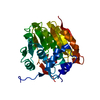
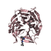
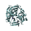
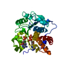
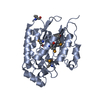
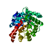
| Title | Strictosidine Synthase from Ophiorrhiza pumila in complex with (S)-1-Ethyl-2,3,4,9-tetrahydro-1H-beta-carboline | ||||||
 Components Components | Strictosidine synthase | ||||||
 Keywords Keywords | LYASE / alkaloid / C-C bond / Pictet-Spenglerase | ||||||
| Function / homology |  Function and homology information Function and homology information | ||||||
| Biological species |  Ophiorrhiza pumila (plant) Ophiorrhiza pumila (plant) | ||||||
| Method |  X-RAY DIFFRACTION / SYNCHROTRON / MOLECULAR REPLACEMENT / Resolution: 2.42 Å X-RAY DIFFRACTION / SYNCHROTRON / MOLECULAR REPLACEMENT / Resolution: 2.42 Å | ||||||
 Authors Authors | Eger, E. / Sharma, M. / Kroutil, W. / Grogan, G. | ||||||
 Citation Citation | Journal: J.Am.Chem.Soc. / Year: 2020 Title: Inverted Binding of Non-natural Substrates in Strictosidine Synthase Leads to a Switch of Stereochemical Outcome in Enzyme-Catalyzed Pictet-Spengler Reactions. Authors: Eger, E. / Simon, A. / Sharma, M. / Yang, S. / Breukelaar, W.B. / Grogan, G. / Houk, K.N. / Kroutil, W. | ||||||
| History |
|
- Structure visualization
| Structure viewer | Molecule: MolmilJmol/JSmol |
|---|
- Downloads & links
-Download
| PDBx/mmCIF format | 6s5j.cif.gz | 74.1 KB | Display | PDBx/mmCIF format |
|---|---|---|---|---|
| PDB format | pdb6s5j.ent.gz | 52.6 KB | Display | PDB format |
| PDBx/mmJSON format | 6s5j.json.gz | Tree view | PDBx/mmJSON format | |
| Others |  Other downloads Other downloads |
-Validation report
| Arichive directory | https://data.pdbj.org/pub/pdb/validation_reports/s5/6s5jftp://data.pdbj.org/pub/pdb/validation_reports/s5/6s5j | HTTPS FTP |
|---|
-Related structure data
| Related structure data | 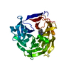 6s5mC  6s5qC 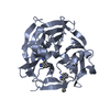 6s5uC 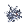 2fp9S S: Starting model for refinement C: citing same article ( |
|---|---|
| Similar structure data |


-Links
 PDBj
PDBj


- Assembly
| Deposited unit | 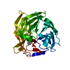
| ||||||||
|---|---|---|---|---|---|---|---|---|---|
| 1 |
| ||||||||
| Unit cell |
|
-Components
| #1: Protein | Mass: 36769.902 Da / Num. of mol.: 1 Source method: isolated from a genetically manipulated source Source: (gene. exp.) Ophiorrhiza pumila (plant) / Gene: str / Plasmid: pET-28a / Production host:  |
|---|---|
| #2: Chemical | ChemComp-KW8 / (  Mass: 200.280 Da / Num. of mol.: 1 / Source method: obtained synthetically / Formula: C13H16N2 / Feature type: SUBJECT OF INVESTIGATION Mass: 200.280 Da / Num. of mol.: 1 / Source method: obtained synthetically / Formula: C13H16N2 / Feature type: SUBJECT OF INVESTIGATION |
| #3: Water | ChemComp-HOH /  Mass: 18.015 Da / Num. of mol.: 13 / Source method: isolated from a natural source / Formula: H2O Mass: 18.015 Da / Num. of mol.: 13 / Source method: isolated from a natural source / Formula: H2O |
| Has ligand of interest | Y |
| Has protein modification | Y |
-Experimental details
-Experiment
| Experiment | Method: X-RAY DIFFRACTION / Number of used crystals: 1 |
|---|
- Sample preparation
| Crystal | Density Matthews: 2.58 Å3/Da / Density % sol: 52.41 % |
|---|---|
| Crystal grow | Temperature: 298 K / Method: vapor diffusion, sitting drop / pH: 8 / Details: 0.1 M Tris-HCl pH 8.0; 0.3 M NH4Cl; 20% PEG 6K |
-Data collection
| Diffraction | Mean temperature: 120 K / Serial crystal experiment: N |
|---|---|
| Diffraction source | Source: SYNCHROTRON / Site: Diamond  / Beamline: I04-1 / Wavelength: 0.92821 Å / Beamline: I04-1 / Wavelength: 0.92821 Å |
| Detector | Type: DECTRIS PILATUS 6M-F / Detector: PIXEL / Date: Nov 28, 2015 |
| Radiation | Protocol: SINGLE WAVELENGTH / Monochromatic (M) / Laue (L): M / Scattering type: x-ray |
| Radiation wavelength | Wavelength: 0.92821 Å / Relative weight: 1 |
| Reflection | Resolution: 2.42→42.01 Å / Num. obs: 14317 / % possible obs: 99.3 % / Redundancy: 6.8 % / CC1/2: 1 / Rmerge(I) obs: 0.05 / Rpim(I) all: 0.03 / Net I/σ(I): 17.2 |
| Reflection shell | Resolution: 2.42→2.51 Å / Redundancy: 7.1 % / Rmerge(I) obs: 0.43 / Mean I/σ(I) obs: 3.5 / Num. unique obs: 1476 / CC1/2: 0.97 / Rpim(I) all: 0.26 / % possible all: 99.3 |
- Processing
| Software |
| ||||||||||||||||||||||||||||||||||||||||||||||||||||||||||||
|---|---|---|---|---|---|---|---|---|---|---|---|---|---|---|---|---|---|---|---|---|---|---|---|---|---|---|---|---|---|---|---|---|---|---|---|---|---|---|---|---|---|---|---|---|---|---|---|---|---|---|---|---|---|---|---|---|---|---|---|---|---|
| Refinement | Method to determine structure: MOLECULAR REPLACEMENT Starting model: 2FP9 Resolution: 2.42→42.01 Å / Cor.coef. Fo:Fc: 0.961 / Cor.coef. Fo:Fc free: 0.936 / SU B: 17.792 / SU ML: 0.336 / Cross valid method: THROUGHOUT / σ(F): 0 / ESU R: 0.399 / ESU R Free: 0.284 Details: HYDROGENS HAVE BEEN ADDED IN THE RIDING POSITIONS U VALUES : REFINED INDIVIDUALLY
| ||||||||||||||||||||||||||||||||||||||||||||||||||||||||||||
| Solvent computation | Ion probe radii: 0.8 Å / Shrinkage radii: 0.8 Å / VDW probe radii: 1.2 Å | ||||||||||||||||||||||||||||||||||||||||||||||||||||||||||||
| Displacement parameters | Biso max: 151.43 Å2 / Biso mean: 65.011 Å2 / Biso min: 30 Å2
| ||||||||||||||||||||||||||||||||||||||||||||||||||||||||||||
| Refinement step | Cycle: final / Resolution: 2.42→42.01 Å
| ||||||||||||||||||||||||||||||||||||||||||||||||||||||||||||
| Refine LS restraints |
| ||||||||||||||||||||||||||||||||||||||||||||||||||||||||||||
| LS refinement shell | Resolution: 2.42→2.483 Å / Rfactor Rfree error: 0 / Total num. of bins used: 20
|
