 Movie
Movie Controller

[English] 日本語
 Yorodumi
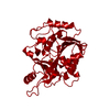
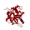
- PDB-1qe5: PURINE NUCLEOSIDE PHOSPHORYLASE FROM CELLULOMONAS SP. IN COMPLEX ... -
+ Open data

- Basic information
| Entry | Database: PDB / ID: 1qe5 | ||||||
|---|---|---|---|---|---|---|---|
| Title | PURINE NUCLEOSIDE PHOSPHORYLASE FROM CELLULOMONAS SP. IN COMPLEX WITH PHOSPHATE | ||||||
 Components Components | PENTOSYLTRANSFERASE | ||||||
 Keywords Keywords | TRANSFERASE / ENZYME / PURINE NUCLEOSIDE PHOSPHORYLASE | ||||||
| Function / homology |  Function and homology information Function and homology informationnucleoside metabolic process / purine-nucleoside phosphorylase activity / purine-nucleoside phosphorylase / cytoplasm Similarity search - Function | ||||||
| Biological species |  Cellulomonas sp. (bacteria) Cellulomonas sp. (bacteria) | ||||||
| Method |  X-RAY DIFFRACTION / SYNCHROTRON / MOLECULAR REPLACEMENT / Resolution: 2.2 Å X-RAY DIFFRACTION / SYNCHROTRON / MOLECULAR REPLACEMENT / Resolution: 2.2 Å | ||||||
 Authors Authors | Tebbe, J. / Bzowska, A. / Wielgus-Kutrowska, B. / Schroeder, W. / Kazimierczuk, Z. / Shugar, D. / Saenger, W. / Koellner, G. | ||||||
 Citation Citation | Journal: J.Mol.Biol. / Year: 1999 Title: Crystal structure of the purine nucleoside phosphorylase (PNP) from Cellulomonas sp. and its implication for the mechanism of trimeric PNPs. Authors: Tebbe, J. / Bzowska, A. / Wielgus-Kutrowska, B. / Schroder, W. / Kazimierczuk, Z. / Shugar, D. / Saenger, W. / Koellner, G. | ||||||
| History |
|
- Structure visualization
| Structure viewer | Molecule: MolmilJmol/JSmol |
|---|
- Downloads & links
-Download
| PDBx/mmCIF format | 1qe5.cif.gz | 159.9 KB | Display | PDBx/mmCIF format |
|---|---|---|---|---|
| PDB format | pdb1qe5.ent.gz | 125 KB | Display | PDB format |
| PDBx/mmJSON format | 1qe5.json.gz | Tree view | PDBx/mmJSON format | |
| Others |  Other downloads Other downloads |
-Validation report
| Arichive directory | https://data.pdbj.org/pub/pdb/validation_reports/qe/1qe5ftp://data.pdbj.org/pub/pdb/validation_reports/qe/1qe5 | HTTPS FTP |
|---|
-Related structure data
| Related structure data | 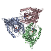 1c3xC 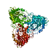 1vfnS S: Starting model for refinement C: citing same article ( |
|---|---|
| Similar structure data |


-Links
 PDBj
PDBj- Assembly
| Deposited unit | 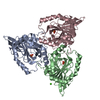
| ||||||||||
|---|---|---|---|---|---|---|---|---|---|---|---|
| 1 |
| ||||||||||
| Unit cell |
| ||||||||||
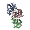
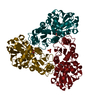
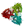
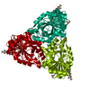
| Details | trimer, constructuted from chain A, B and C through a three-fold non- crystallographic symmetry axis |
-Components
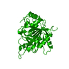
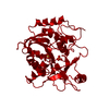
| #1: Protein | Mass: 27546.156 Da / Num. of mol.: 3 / Fragment: RESIDUES 9-282 / Source method: isolated from a natural source / Source: (natural) Cellulomonas sp. (bacteria)References: UniProt: P81989, purine-nucleoside phosphorylase #2: Chemical |   Mass: 94.971 Da / Num. of mol.: 3 / Source method: obtained synthetically / Formula: PO4 Mass: 94.971 Da / Num. of mol.: 3 / Source method: obtained synthetically / Formula: PO4#3: Chemical | ChemComp-CA / |   Mass: 40.078 Da / Num. of mol.: 1 / Source method: obtained synthetically / Formula: Ca Mass: 40.078 Da / Num. of mol.: 1 / Source method: obtained synthetically / Formula: Ca#4: Water | ChemComp-HOH / |  Mass: 18.015 Da / Num. of mol.: 262 / Source method: isolated from a natural source / Formula: H2O Mass: 18.015 Da / Num. of mol.: 262 / Source method: isolated from a natural source / Formula: H2O |
|---|
-Experimental details
-Experiment
| Experiment | Method: X-RAY DIFFRACTION / Number of used crystals: 1 |
|---|
- Sample preparation
| Crystal | Density Matthews: 2.52 Å3/Da / Density % sol: 51.16 % | ||||||||||||||||||||||||||||||
|---|---|---|---|---|---|---|---|---|---|---|---|---|---|---|---|---|---|---|---|---|---|---|---|---|---|---|---|---|---|---|---|
| Crystal grow | Temperature: 298 K / Method: vapor diffusion, hanging drop Details: PEG 4000, cacodylate, Ca-acetate, sodium phosphate, VAPOR DIFFUSION, HANGING DROP, temperature 298K | ||||||||||||||||||||||||||||||
| Crystal grow | *PLUS pH: 7 / Method: unknown | ||||||||||||||||||||||||||||||
| Components of the solutions | *PLUS
|
-Data collection
| Diffraction | Mean temperature: 277 K |
|---|---|
| Diffraction source | Source: SYNCHROTRON / Site: EMBL/DESY, HAMBURG  / Beamline: X11 / Wavelength: 0.92 / Beamline: X11 / Wavelength: 0.92 |
| Detector | Type: MARRESEARCH / Detector: IMAGE PLATE / Date: Oct 15, 1997 |
| Radiation | Protocol: SINGLE WAVELENGTH / Monochromatic (M) / Laue (L): M / Scattering type: x-ray |
| Radiation wavelength | Wavelength: 0.92 Å / Relative weight: 1 |
| Reflection | Resolution: 2.2→32 Å / Num. all: 43153 / Num. obs: 37330 / % possible obs: 86 % / Observed criterion σ(F): 0 / Observed criterion σ(I): 0 / Redundancy: 4.4 % / Biso Wilson estimate: 21.8 Å2 / Rmerge(I) obs: 0.075 / Net I/σ(I): 13.3 |
| Reflection shell | Resolution: 2.2→2.24 Å / Rmerge(I) obs: 0.299 / % possible all: 79.5 |
| Reflection shell | *PLUS % possible obs: 80 % |
- Processing
| Software |
| ||||||||||||||||||||||||||||||||||||
|---|---|---|---|---|---|---|---|---|---|---|---|---|---|---|---|---|---|---|---|---|---|---|---|---|---|---|---|---|---|---|---|---|---|---|---|---|---|
| Refinement | Method to determine structure: MOLECULAR REPLACEMENT Starting model: 1VFN Resolution: 2.2→28 Å / Cross valid method: THROUGHOUT / σ(F): 0 / σ(I): 0 / Stereochemistry target values: Engh & Huber
| ||||||||||||||||||||||||||||||||||||
| Solvent computation | Bsol: 45.88 Å2 / ksol: 0.327 e/Å3 | ||||||||||||||||||||||||||||||||||||
| Displacement parameters | Biso mean: 22.94 Å2 | ||||||||||||||||||||||||||||||||||||
| Refine analyze |
| ||||||||||||||||||||||||||||||||||||
| Refinement step | Cycle: LAST / Resolution: 2.2→28 Å
| ||||||||||||||||||||||||||||||||||||
| Refine LS restraints |
| ||||||||||||||||||||||||||||||||||||
| LS refinement shell | Resolution: 2.2→2.28 Å / Total num. of bins used: 10
| ||||||||||||||||||||||||||||||||||||
| Software | *PLUS Name: 'CNS' / Classification: refinement | ||||||||||||||||||||||||||||||||||||
| Refine LS restraints | *PLUS
|
