 Movie
Movie Controller


+ Open data

- Basic information
| Entry | Database: PDB / ID: 1vfn | ||||||
|---|---|---|---|---|---|---|---|
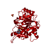
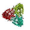
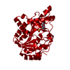
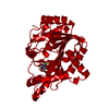
| Title | PURINE NUCLEOSIDE PHOSPHORYLASE | ||||||
 Components Components | PURINE-NUCLEOSIDE PHOSPHORYLASE | ||||||
 Keywords Keywords | NUCLEOSIDE PHOSPHORYLASE / TRANSFERASE / GLYCOSYLTRANSFERASE / DISEASE MUTATION | ||||||
| Function / homology |  Function and homology information Function and homology informationguanosine phosphorylase activity / purine-nucleoside phosphorylase / purine-nucleoside phosphorylase activity / purine ribonucleoside salvage / cytoplasm Similarity search - Function | ||||||
| Biological species |  | ||||||
| Method |  X-RAY DIFFRACTION / SYNCHROTRON / MOLECULAR REPLACEMENT / Resolution: 2.15 Å X-RAY DIFFRACTION / SYNCHROTRON / MOLECULAR REPLACEMENT / Resolution: 2.15 Å | ||||||
 Authors Authors | Koellner, G. / Bzowska, A. | ||||||
 Citation Citation | Journal: J.Mol.Biol. / Year: 1997 Title: Crystal structure of calf spleen purine nucleoside phosphorylase in a complex with hypoxanthine at 2.15 A resolution. Authors: Koellner, G. / Luic, M. / Shugar, D. / Saenger, W. / Bzowska, A. #1: Journal: FEBS Lett. / Year: 1995Title: Calf Spleen Purine Nucleoside Phosphorylase: Purification, Sequence and Crystal Structure of its Complex with an N(7)-Acycloguanosine Inhibitor Authors: Bzowska, A. / Luic, M. / Schroder, W. / Shugar, D. / Saenger, W. / Koellner, G. | ||||||
| History |
|
- Structure visualization
| Structure viewer | Molecule: MolmilJmol/JSmol |
|---|
- Downloads & links
-Download
| PDBx/mmCIF format | 1vfn.cif.gz | 70.4 KB | Display | PDBx/mmCIF format |
|---|---|---|---|---|
| PDB format | pdb1vfn.ent.gz | 50.9 KB | Display | PDB format |
| PDBx/mmJSON format | 1vfn.json.gz | Tree view | PDBx/mmJSON format | |
| Others |  Other downloads Other downloads |
-Validation report
| Arichive directory | https://data.pdbj.org/pub/pdb/validation_reports/vf/1vfnftp://data.pdbj.org/pub/pdb/validation_reports/vf/1vfn | HTTPS FTP |
|---|
-Related structure data
| Related structure data | 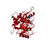 1ulbS S: Starting model for refinement |
|---|---|
| Similar structure data |


-Links
 PDBj
PDBj
- Assembly
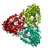
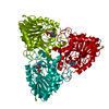
| Deposited unit | 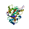
| ||||||||||||
|---|---|---|---|---|---|---|---|---|---|---|---|---|---|
| 1 | 
| ||||||||||||
| 2 | 
| ||||||||||||
| Unit cell |
| ||||||||||||
| Components on special symmetry positions |
|
-Components
| #1: Protein | Mass: 31312.701 Da / Num. of mol.: 1 / Source method: isolated from a natural source / Source: (natural) References: UniProt: P55859, purine-nucleoside phosphorylase |
|---|---|
| #2: Chemical | ChemComp-MG /   Mass: 24.305 Da / Num. of mol.: 1 / Source method: obtained synthetically / Formula: Mg Mass: 24.305 Da / Num. of mol.: 1 / Source method: obtained synthetically / Formula: Mg |
| #3: Chemical | ChemComp-ZN /   Mass: 65.409 Da / Num. of mol.: 1 / Source method: obtained synthetically / Formula: Zn Mass: 65.409 Da / Num. of mol.: 1 / Source method: obtained synthetically / Formula: Zn |
| #4: Chemical | ChemComp-HPA /   Mass: 136.111 Da / Num. of mol.: 1 / Source method: obtained synthetically / Formula: C5H4N4O Mass: 136.111 Da / Num. of mol.: 1 / Source method: obtained synthetically / Formula: C5H4N4O |
| #5: Water | ChemComp-HOH /  Mass: 18.015 Da / Num. of mol.: 129 / Source method: isolated from a natural source / Formula: H2O Mass: 18.015 Da / Num. of mol.: 129 / Source method: isolated from a natural source / Formula: H2O |
-Experimental details
-Experiment
| Experiment | Method: X-RAY DIFFRACTION / Number of used crystals: 1 |
|---|
- Sample preparation
| Crystal | Density Matthews: 2.22 Å3/Da / Density % sol: 43 % | ||||||||||||||||||||||||||||||||||||||||||||||||||||||||||||
|---|---|---|---|---|---|---|---|---|---|---|---|---|---|---|---|---|---|---|---|---|---|---|---|---|---|---|---|---|---|---|---|---|---|---|---|---|---|---|---|---|---|---|---|---|---|---|---|---|---|---|---|---|---|---|---|---|---|---|---|---|---|
| Crystal grow | pH: 8 Details: 16%(W/V) PEG 4000, 0.04M TRIS-HCL BUFFER PH 8.2-8.5, 0.08M MGCL2, pH 8.0 PH range: 8.2-8.5 | ||||||||||||||||||||||||||||||||||||||||||||||||||||||||||||
| Crystal grow | *PLUS Temperature: 18 ℃ / Method: vapor diffusion, hanging drop | ||||||||||||||||||||||||||||||||||||||||||||||||||||||||||||
| Components of the solutions | *PLUS
|
-Data collection
| Diffraction | Mean temperature: 277 K |
|---|---|
| Diffraction source | Source: SYNCHROTRON / Site: EMBL/DESY, HAMBURG  / Beamline: X31 / Wavelength: 0.9204 / Beamline: X31 / Wavelength: 0.9204 |
| Detector | Type: MARRESEARCH / Detector: IMAGE PLATE / Date: Mar 1, 1996 |
| Radiation | Monochromatic (M) / Laue (L): M / Scattering type: x-ray |
| Radiation wavelength | Wavelength: 0.9204 Å / Relative weight: 1 |
| Reflection | Resolution: 2.15→20 Å / Num. obs: 15158 / % possible obs: 98.4 % / Observed criterion σ(I): 1 / Redundancy: 3.27 % / Rmerge(I) obs: 0.074 / Net I/σ(I): 10.6 |
| Reflection shell | Resolution: 2.1→2.2 Å / Rmerge(I) obs: 0.212 / Mean I/σ(I) obs: 6 / % possible all: 97.6 |
- Processing
| Software |
| ||||||||||||||||||||||||||||||||||||||||||||||||||
|---|---|---|---|---|---|---|---|---|---|---|---|---|---|---|---|---|---|---|---|---|---|---|---|---|---|---|---|---|---|---|---|---|---|---|---|---|---|---|---|---|---|---|---|---|---|---|---|---|---|---|---|
| Refinement | Method to determine structure: MOLECULAR REPLACEMENT Starting model: PDB ENTRY 1ULB Resolution: 2.15→20 Å / Isotropic thermal model: TNT PACKAGE / σ(F): 1 / Stereochemistry target values: TNT PACKAGE
| ||||||||||||||||||||||||||||||||||||||||||||||||||
| Solvent computation | Solvent model: TNT PACKAGE / Bsol: 173 Å2 / ksol: 0.665 e/Å3 | ||||||||||||||||||||||||||||||||||||||||||||||||||
| Refinement step | Cycle: LAST / Resolution: 2.15→20 Å
| ||||||||||||||||||||||||||||||||||||||||||||||||||
| Refine LS restraints |
| ||||||||||||||||||||||||||||||||||||||||||||||||||
| Software | *PLUS Name: TNT / Classification: refinement | ||||||||||||||||||||||||||||||||||||||||||||||||||
| Refinement | *PLUS Rfactor obs: 0.18 | ||||||||||||||||||||||||||||||||||||||||||||||||||
| Solvent computation | *PLUS | ||||||||||||||||||||||||||||||||||||||||||||||||||
| Displacement parameters | *PLUS | ||||||||||||||||||||||||||||||||||||||||||||||||||
| Refine LS restraints | *PLUS
|
