 Movie
Movie Controller


+ Open data

- Basic information
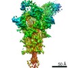
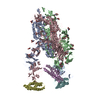
| Entry | Database: PDB / ID: 1qd7 | |||||||||
|---|---|---|---|---|---|---|---|---|---|---|
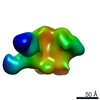
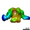
| Title | PARTIAL MODEL FOR 30S RIBOSOMAL SUBUNIT | |||||||||
 Components Components |
| |||||||||
 Keywords Keywords | RIBOSOME / 30S RIBOSOMAL SUBUNIT / LOW RESOLUTION MODEL | |||||||||
| Function / homology |  Function and homology information Function and homology informationribosome biogenesis / ribosomal small subunit biogenesis / small ribosomal subunit / small ribosomal subunit rRNA binding / cytosolic small ribosomal subunit / tRNA binding / rRNA binding / structural constituent of ribosome / ribosome / translation ...ribosome biogenesis / ribosomal small subunit biogenesis / small ribosomal subunit / small ribosomal subunit rRNA binding / cytosolic small ribosomal subunit / tRNA binding / rRNA binding / structural constituent of ribosome / ribosome / translation / ribonucleoprotein complex / response to antibiotic / cytoplasm Similarity search - Function | |||||||||
| Biological species |   Thermus thermophilus (bacteria) Thermus thermophilus (bacteria) | |||||||||
| Method |  X-RAY DIFFRACTION / SYNCHROTRON / MAD / Resolution: 5.5 Å X-RAY DIFFRACTION / SYNCHROTRON / MAD / Resolution: 5.5 Å | |||||||||
 Authors Authors | Clemons Jr., W.M. / May, J.L.C. / Wimberly, B.T. / McCutcheon, J.P. / Capel, M.S. / Ramakrishnan, V. | |||||||||
 Citation Citation | Journal: Nature / Year: 1999 Title: Structure of a bacterial 30S ribosomal subunit at 5.5 A resolution. Authors: Clemons Jr., W.M. / May, J.L. / Wimberly, B.T. / McCutcheon, J.P. / Capel, M.S. / Ramakrishnan, V. #1: Journal: Embo J. / Year: 1998Title: The crystal structure of ribosomal protein S4 reveals a two-domain molecule with an extensive RNA-binding surface: one domain shows structural homology to the ETS DNA-binding motif Authors: Davies, C. / Gerstner, R.B. / Draper, D.E. / Ramakrishnan, V. / White, S.W. #2: Journal: Nature / Year: 1992Title: The structure of ribosomal protein S5 reveals sites of interaction with 16S rRNA Authors: Ramakrishnan, V. / White, S.W. #3: Journal: Embo J. / Year: 1994Title: Crystal structure of the ribosomal protein S6 from Thermus thermophilus Authors: Lindahl, M. / Svensson, L.A. / Liljas, A. / Sedelnikova, I.A. / Eliseikina, I.A. / Fomenkova, N.P. / Nevskaya, N. / Nikonov, S.V. / Garber, M.B. / Muranova, T.A. / Rykonova, A.I. / Amons, R. #4: Journal: Structure / Year: 1997Title: The structure of ribosomal protein S7 at 1.9 A resolution reveals a beta- hairpin motif that binds double-stranded nucleic acids Authors: Wimberly, B.T. / White, S.W. / Ramakrishnan, V. #5: Journal: J.Mol.Biol. / Year: 1998Title: Crystal structure of ribosomal protein S8 from Thermus thermophilus reveals a high degree of structural conservation of a specific RNA binding site Authors: Nevskaya, N. / Tischenko, S. / Nikulin, A. / Al-Karadaghi, S. / Liljas, A. / Ehresmann, B. / Ehresmann, C. / Garber, M. / Nikonov, S. #6: Journal: Structure / Year: 1998Title: Conformational variability of the N-terminal helix in the structure of ribosomal protein S15 Authors: Clemons Jr., W.M. / Davies, C.R. / White, S.W. / Ramakrishnan, V. #7: Journal: Biochemistry / Year: 1996Title: Solution structure of prokaryotic ribosomal protein S17 by high-resolution NMR spectroscopy Authors: Jaishree, T.N. / Ramakrishnan, V. / White, S.W. | |||||||||
| History |
|
- Structure visualization
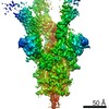
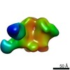
| Structure viewer | Molecule: MolmilJmol/JSmol |
|---|
- Downloads & links
-Download
| PDBx/mmCIF format | 1qd7.cif.gz | 60.8 KB | Display | PDBx/mmCIF format |
|---|---|---|---|---|
| PDB format | pdb1qd7.ent.gz | 33.8 KB | Display | PDB format |
| PDBx/mmJSON format | 1qd7.json.gz | Tree view | PDBx/mmJSON format | |
| Others |  Other downloads Other downloads |
-Validation report
| Arichive directory | https://data.pdbj.org/pub/pdb/validation_reports/qd/1qd7ftp://data.pdbj.org/pub/pdb/validation_reports/qd/1qd7 | HTTPS FTP |
|---|
-Related structure data
| Related structure data |  1a23S 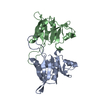 1an7S  1pkpS  1risS 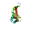 1rssS S: Starting model for refinement |
|---|---|
| Similar structure data |

-Links
 PDBj
PDBj


- Assembly
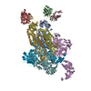
| Deposited unit | 
| ||||||||||
|---|---|---|---|---|---|---|---|---|---|---|---|
| 1 |
| ||||||||||
| Unit cell |
|
-Components
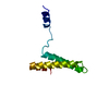
-RNA chain , 2 types, 2 molecules AB
| #1: RNA chain | Mass: 53096.625 Da / Num. of mol.: 1 / Fragment: RESIDUES 563-912 / Source method: isolated from a natural source / Source: (natural) Thermus thermophilus (bacteria) |
|---|---|
| #2: RNA chain | Mass: 17211.391 Da / Num. of mol.: 1 / Fragment: RESIDUES 1400-1500 / Source method: isolated from a natural source / Source: (natural) Thermus thermophilus (bacteria) |
-Protein , 8 types, 8 molecules CDEFGHIJ
| #3: Protein | Mass: 18624.359 Da / Num. of mol.: 1 / Source method: isolated from a natural source / Source: (natural) Thermus thermophilus (bacteria) / References: UniProt: P81288 |
|---|---|
| #4: Protein | Mass: 15240.724 Da / Num. of mol.: 1 / Source method: isolated from a natural source / Source: (natural) Thermus thermophilus (bacteria) / References: UniProt: P02357 |
| #5: Protein | Mass: 11619.337 Da / Num. of mol.: 1 / Source method: isolated from a natural source / Source: (natural) Thermus thermophilus (bacteria) / References: UniProt: P23370, UniProt: Q5SLP8*PLUS |
| #6: Protein | Mass: 15342.846 Da / Num. of mol.: 1 / Source method: isolated from a natural source / Source: (natural) Thermus thermophilus (bacteria) / References: UniProt: P17291 |
| #7: Protein | Mass: 15624.214 Da / Num. of mol.: 1 / Source method: isolated from a natural source / Source: (natural) Thermus thermophilus (bacteria) / References: UniProt: P24319, UniProt: P0DOY9*PLUS |
| #8: Protein | Mass: 10200.844 Da / Num. of mol.: 1 / Source method: isolated from a natural source / Source: (natural) Thermus thermophilus (bacteria) / References: UniProt: P05766 |
| #9: Protein | Mass: 10081.853 Da / Num. of mol.: 1 / Source method: isolated from a natural source / Source: (natural) Thermus thermophilus (bacteria) / References: UniProt: P23828 |
| #10: Protein | Mass: 8528.504 Da / Num. of mol.: 1 / Source method: isolated from a natural source / Source: (natural) Thermus thermophilus (bacteria) |
-Experimental details
-Experiment
| Experiment | Method: X-RAY DIFFRACTION / Number of used crystals: 6 |
|---|
- Sample preparation
| Crystal grow | Temperature: 277 K / Method: vapor diffusion / Details: VAPOUR DIFFUSION AT 277 K, MPD, VAPOR DIFFUSION | ||||||||||||
|---|---|---|---|---|---|---|---|---|---|---|---|---|---|
| Components of the solutions |
| ||||||||||||
| Crystal grow | *PLUS Method: vapor diffusion, hanging drop / Details: Trakhanov, S.D., (1987) FEBS Lett., 220, 319. | ||||||||||||
| Components of the solutions | *PLUS
|
-Data collection
| Diffraction | Mean temperature: 100 K | |||||||||
|---|---|---|---|---|---|---|---|---|---|---|
| Diffraction source | Source: SYNCHROTRON / Site: NSLS  / Beamline: X25 / Wavelength: 1.000, 1.700 / Beamline: X25 / Wavelength: 1.000, 1.700 | |||||||||
| Detector | Type: BRANDEIS - B4 / Detector: CCD / Date: Mar 4, 1999 | |||||||||
| Radiation | Protocol: SINGLE WAVELENGTH / Monochromatic (M) / Laue (L): M / Scattering type: x-ray | |||||||||
| Radiation wavelength |
| |||||||||
| Reflection | Highest resolution: 5.5 Å / Num. all: 42000 / Num. obs: 42000 / % possible obs: 95 % / Observed criterion σ(I): 0 / Redundancy: 5 % | |||||||||
| Reflection shell | Highest resolution: 5.5 Å / Mean I/σ(I) obs: 5.1 / % possible all: 83 | |||||||||
| Reflection | *PLUS |
- Processing
| Software |
| ||||||||||||
|---|---|---|---|---|---|---|---|---|---|---|---|---|---|
| Refinement | Method to determine structure: MAD Starting model: PROTEINS WERE FIT INTO MAP AS RIGID BODIES FROM KNOWN CRYSTAL OR NMR STRUCTURES OF ISOLATED PROTEINS EXCEPT FOR S20, FOR WHICH NO HIGH RESOLUTION STRUCTURE EXISTS. S20 WAS FIT AS ...Starting model: PROTEINS WERE FIT INTO MAP AS RIGID BODIES FROM KNOWN CRYSTAL OR NMR STRUCTURES OF ISOLATED PROTEINS EXCEPT FOR S20, FOR WHICH NO HIGH RESOLUTION STRUCTURE EXISTS. S20 WAS FIT AS THREE-HELIX BUNDLE.S4 SEE REFERENCE (1); S5 MODELED ACCORDING TO PDB CODE 1PKP;S6 MODELED ACCORDING TO PDB CODE 1RIS;S7 MODELED ACCORDING TO PDB CODE 1RSS;S8 MODELED ACCORDING TO PDB CODE 1AN7;S15 MODELED ACCORDING TO PDB CODE 1A23;S17 SEE REFERENCE (7); Highest resolution: 5.5 Å / Num. reflection all: 42000 / Num. reflection obs: 42000 Details: NO REFINEMENT WAS DONE EXCEPT FOR VISUAL MAP FITTING. THIS MODEL WAS MADE FROM A 5.5 ANGSTROM X-RAY MAP BY FITTING A-FORM RNA HELICES INTO REGIONS OF DOUBLE- HELICAL DENSITY. NO ATTEMPT HAS ...Details: NO REFINEMENT WAS DONE EXCEPT FOR VISUAL MAP FITTING. THIS MODEL WAS MADE FROM A 5.5 ANGSTROM X-RAY MAP BY FITTING A-FORM RNA HELICES INTO REGIONS OF DOUBLE- HELICAL DENSITY. NO ATTEMPT HAS BEEN MADE TO MAINTAIN PROPER STEREOCHEMISTRY OR EVEN PHOSPHATE-PHOSPHATE DISTANCES. IN PARTICULAR AT JUNCTIONS OF THE SHORT HELICES, THE CHAIN MAY BE UNREALISTIC | ||||||||||||
| Refinement step | Cycle: LAST / Highest resolution: 5.5 Å
| ||||||||||||
| Refinement | *PLUS Highest resolution: 5.5 Å | ||||||||||||
| Solvent computation | *PLUS | ||||||||||||
| Displacement parameters | *PLUS |
