 Movie
Movie Controller

[English] 日本語
 Yorodumi
- PDB-1qd2: CRYSTAL STRUCTURE OF THE COMPLEX OF TRICHOSANTHIN WITH ADENINE, O... -
+ Open data

- Basic information
| Entry | Database: PDB / ID: 1qd2 | ||||||
|---|---|---|---|---|---|---|---|
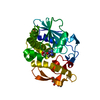
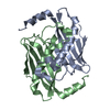
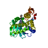
| Title | CRYSTAL STRUCTURE OF THE COMPLEX OF TRICHOSANTHIN WITH ADENINE, OBTAINED FROM TRICHOSANTHIN COMPLEXED WITH THE DINUCLEOTIDE APG | ||||||
 Components Components | TRICHOSANTHIN | ||||||
 Keywords Keywords | HYDROLASE / ENZYME-PRODUCT COMPLEX OBTAINED FROM ENZYME-SUBSTRATE ANALOG COMPLEX | ||||||
| Function / homology |  Function and homology information Function and homology informationregulation of defense response to virus / rRNA N-glycosylase / rRNA N-glycosylase activity / defense response / toxin activity / negative regulation of translation Similarity search - Function | ||||||
| Biological species |  Trichosanthes kirilowii (Chinese cucumber) Trichosanthes kirilowii (Chinese cucumber) | ||||||
| Method |  X-RAY DIFFRACTION / Resolution: 1.86 Å X-RAY DIFFRACTION / Resolution: 1.86 Å | ||||||
 Authors Authors | Gu, Y.J. / Xia, Z.X. | ||||||
 Citation Citation | Journal: Proteins / Year: 2000 Title: Crystal structures of the complexes of trichosanthin with four substrate analogs and catalytic mechanism of RNA N-glycosidase. Authors: Gu, Y.J. / Xia, Z.X. | ||||||
| History |
|
- Structure visualization
| Structure viewer | Molecule: MolmilJmol/JSmol |
|---|
- Downloads & links
-Download
| PDBx/mmCIF format | 1qd2.cif.gz | 63.2 KB | Display | PDBx/mmCIF format |
|---|---|---|---|---|
| PDB format | pdb1qd2.ent.gz | 46.5 KB | Display | PDB format |
| PDBx/mmJSON format | 1qd2.json.gz | Tree view | PDBx/mmJSON format | |
| Others |  Other downloads Other downloads |
-Validation report
| Arichive directory | https://data.pdbj.org/pub/pdb/validation_reports/qd/1qd2ftp://data.pdbj.org/pub/pdb/validation_reports/qd/1qd2 | HTTPS FTP |
|---|
-Related structure data
| Related structure data | |
|---|---|
| Similar structure data |


-Links
 PDBj
PDBj


- Assembly
| Deposited unit | 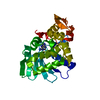
| ||||||||||
|---|---|---|---|---|---|---|---|---|---|---|---|
| 1 |
| ||||||||||
| Unit cell |
|
-Components
| #1: Protein | Mass: 27166.770 Da / Num. of mol.: 1 / Source method: isolated from a natural source Source: (natural) Trichosanthes kirilowii (Chinese cucumber)References: UniProt: P09989, rRNA N-glycosylase |
|---|---|
| #2: Chemical | ChemComp-ADE /   Mass: 135.127 Da / Num. of mol.: 1 / Source method: obtained synthetically / Formula: C5H5N5 Mass: 135.127 Da / Num. of mol.: 1 / Source method: obtained synthetically / Formula: C5H5N5 |
| #3: Water | ChemComp-HOH /  Mass: 18.015 Da / Num. of mol.: 189 / Source method: isolated from a natural source / Formula: H2O Mass: 18.015 Da / Num. of mol.: 189 / Source method: isolated from a natural source / Formula: H2O |
-Experimental details
-Experiment
| Experiment | Method: X-RAY DIFFRACTION / Number of used crystals: 1 |
|---|
- Sample preparation
| Crystal | Density Matthews: 2.15 Å3/Da / Density % sol: 42.89 % | ||||||||||||||||||||||||||||||||||||||||||
|---|---|---|---|---|---|---|---|---|---|---|---|---|---|---|---|---|---|---|---|---|---|---|---|---|---|---|---|---|---|---|---|---|---|---|---|---|---|---|---|---|---|---|---|
| Crystal grow | Temperature: 281 K / Method: vapor diffusion, hanging drop / pH: 5 Details: HANGING DROP, CITRATE BUFFER, POTASSIUM CHLORIDE, DINUCLEOTIDE APG. NATIVE CRYSTALS SOAKED IN SOLUTION CONTAINING DINUCLEOTIDE APG, pH 5.0, VAPOR DIFFUSION, HANGING DROP, temperature 281K | ||||||||||||||||||||||||||||||||||||||||||
| Crystal grow | *PLUS Temperature: 20 ℃ | ||||||||||||||||||||||||||||||||||||||||||
| Components of the solutions | *PLUS
|
-Data collection
| Diffraction | Mean temperature: 293 K |
|---|---|
| Diffraction source | Source: SEALED TUBE / Wavelength: 1.5418 |
| Detector | Type: MARRESEARCH / Detector: IMAGE PLATE / Date: Jun 15, 1997 |
| Radiation | Protocol: SINGLE WAVELENGTH / Monochromatic (M) / Laue (L): M / Scattering type: x-ray |
| Radiation wavelength | Wavelength: 1.5418 Å / Relative weight: 1 |
| Reflection | Resolution: 1.86→10 Å / Num. all: 20045 / Num. obs: 20045 / % possible obs: 98.8 % / Observed criterion σ(I): 0 / Redundancy: 3.5 % / Biso Wilson estimate: 11.25 Å2 / Rmerge(I) obs: 0.078 / Net I/σ(I): 20 |
| Reflection shell | Resolution: 1.86→1.9 Å / Redundancy: 3 % / Rmerge(I) obs: 0.343 / % possible all: 90.3 |
| Reflection | *PLUS |
| Reflection shell | *PLUS % possible obs: 90.3 % |
- Processing
| Software |
| ||||||||||||||||||||||||||||||||||||||||||||||||||||||||||||
|---|---|---|---|---|---|---|---|---|---|---|---|---|---|---|---|---|---|---|---|---|---|---|---|---|---|---|---|---|---|---|---|---|---|---|---|---|---|---|---|---|---|---|---|---|---|---|---|---|---|---|---|---|---|---|---|---|---|---|---|---|---|
| Refinement | Resolution: 1.86→10 Å / σ(F): 2 / Stereochemistry target values: X-PLOR DICTIONARY
| ||||||||||||||||||||||||||||||||||||||||||||||||||||||||||||
| Refinement step | Cycle: LAST / Resolution: 1.86→10 Å
| ||||||||||||||||||||||||||||||||||||||||||||||||||||||||||||
| Refine LS restraints |
| ||||||||||||||||||||||||||||||||||||||||||||||||||||||||||||
| Software | *PLUS Name: X-PLOR / Version: 3.1 / Classification: refinement | ||||||||||||||||||||||||||||||||||||||||||||||||||||||||||||
| Refinement | *PLUS Lowest resolution: 10 Å / σ(F): 2 / % reflection Rfree: 10 % | ||||||||||||||||||||||||||||||||||||||||||||||||||||||||||||
| Solvent computation | *PLUS | ||||||||||||||||||||||||||||||||||||||||||||||||||||||||||||
| Displacement parameters | *PLUS |
