 Movie
Movie Controller


+ Open data

- Basic information
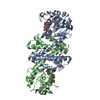
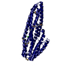
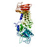
| Entry | Database: PDB / ID: 1qbz | ||||||
|---|---|---|---|---|---|---|---|
| Title | THE CRYSTAL STRUCTURE OF THE SIV GP41 ECTODOMAIN AT 1.47 A | ||||||
 Components Components | PROTEIN (SIV GP41 ECTODOMAIN) | ||||||
 Keywords Keywords | ENVELOPE GLYCOPROTEIN / SIV / HIV / GP41 / MEMBRANE FUSION / PEPTIDE INHIBITOR / SIV ENVELOPE GLYCOPROTEIN GP41 | ||||||
| Function / homology |  Function and homology information Function and homology informationmembrane fusion involved in viral entry into host cell / host cell endosome membrane / viral envelope / symbiont entry into host cell / virion attachment to host cell / host cell plasma membrane / virion membrane / structural molecule activity Similarity search - Function | ||||||
| Biological species |  Simian immunodeficiency virus Simian immunodeficiency virus | ||||||
| Method |  X-RAY DIFFRACTION / SYNCHROTRON / MIR, molecular replacement / Resolution: 1.47 Å X-RAY DIFFRACTION / SYNCHROTRON / MIR, molecular replacement / Resolution: 1.47 Å | ||||||
 Authors Authors | Yang, Z.-N. / Mueser, T.C. / Kaufman, J. / Stahl, S.J. / Wingfield, P.T. / Hyde, C.C. | ||||||
 Citation Citation | Journal: J.Struct.Biol. / Year: 1999 Title: The crystal structure of the SIV gp41 ectodomain at 1.47 A resolution. Authors: Yang, Z.N. / Mueser, T.C. / Kaufman, J. / Stahl, S.J. / Wingfield, P.T. / Hyde, C.C. | ||||||
| History |
|
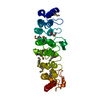
- Structure visualization
| Structure viewer | Molecule: MolmilJmol/JSmol |
|---|
- Downloads & links
-Download
| PDBx/mmCIF format | 1qbz.cif.gz | 167 KB | Display | PDBx/mmCIF format |
|---|---|---|---|---|
| PDB format | pdb1qbz.ent.gz | 135.5 KB | Display | PDB format |
| PDBx/mmJSON format | 1qbz.json.gz | Tree view | PDBx/mmJSON format | |
| Others |  Other downloads Other downloads |
-Validation report
| Arichive directory | https://data.pdbj.org/pub/pdb/validation_reports/qb/1qbzftp://data.pdbj.org/pub/pdb/validation_reports/qb/1qbz | HTTPS FTP |
|---|
-Related structure data
| Related structure data | 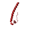 1mofS S: Starting model for refinement |
|---|---|
| Similar structure data |


-Links
 PDBj
PDBj


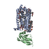
- Assembly
| Deposited unit | 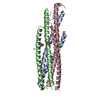
| ||||||||||||
|---|---|---|---|---|---|---|---|---|---|---|---|---|---|
| 1 |
| ||||||||||||
| 2 | 
| ||||||||||||
| Unit cell |
| ||||||||||||
| Components on special symmetry positions |
|
-Components
| #1: Protein | Mass: 14431.267 Da / Num. of mol.: 3 / Fragment: SIV GP41 ECTODOMAIN 27-149 / Mutation: C86A, C92A Source method: isolated from a genetically manipulated source Source: (gene. exp.) Simian immunodeficiency virus / Genus: Lentivirus / Strain: MAC239 / Cellular location: VIRAL MEMBRANE / Production host:  #2: Chemical |   Mass: 200.590 Da / Num. of mol.: 2 / Source method: obtained synthetically / Formula: Hg Mass: 200.590 Da / Num. of mol.: 2 / Source method: obtained synthetically / Formula: Hg#3: Chemical |   Mass: 118.174 Da / Num. of mol.: 2 / Source method: obtained synthetically / Formula: C6H14O2 / Comment: precipitant*YM Mass: 118.174 Da / Num. of mol.: 2 / Source method: obtained synthetically / Formula: C6H14O2 / Comment: precipitant*YM#4: Chemical |   Mass: 35.453 Da / Num. of mol.: 3 / Source method: obtained synthetically / Formula: Cl Mass: 35.453 Da / Num. of mol.: 3 / Source method: obtained synthetically / Formula: Cl#5: Water | ChemComp-HOH / |  Mass: 18.015 Da / Num. of mol.: 263 / Source method: isolated from a natural source / Formula: H2O Mass: 18.015 Da / Num. of mol.: 263 / Source method: isolated from a natural source / Formula: H2O |
|---|
-Experimental details
-Experiment
| Experiment | Method: X-RAY DIFFRACTION / Number of used crystals: 1 |
|---|
- Sample preparation
| Crystal | Density Matthews: 1.78 Å3/Da / Density % sol: 30.8 % | ||||||||||||||||||||||||||||||||||||
|---|---|---|---|---|---|---|---|---|---|---|---|---|---|---|---|---|---|---|---|---|---|---|---|---|---|---|---|---|---|---|---|---|---|---|---|---|---|
| Crystal grow | pH: 4.25 Details: 14-25% MPD (2-METHYL-2,4- PENTANEDIOL) 140-220 MM NACL, 20-40 MM SODIUM ACETATE, pH 4.25 | ||||||||||||||||||||||||||||||||||||
| Crystal grow | *PLUS pH: 3 / Method: vapor diffusion, sitting drop | ||||||||||||||||||||||||||||||||||||
| Components of the solutions | *PLUS
|
-Data collection
| Diffraction | Mean temperature: 93 K |
|---|---|
| Diffraction source | Source: SYNCHROTRON / Site: NSLS  / Beamline: X9B / Wavelength: 0.97564 / Beamline: X9B / Wavelength: 0.97564 |
| Detector | Type: MARRESEARCH / Detector: IMAGE PLATE / Date: Nov 22, 1997 |
| Radiation | Protocol: SINGLE WAVELENGTH / Monochromatic (M) / Laue (L): M / Scattering type: x-ray |
| Radiation wavelength | Wavelength: 0.97564 Å / Relative weight: 1 |
| Reflection | Resolution: 1.47→15 Å / Num. obs: 49553 / % possible obs: 89.9 % / Observed criterion σ(I): 0.5 / Redundancy: 8.3 % / Rmerge(I) obs: 0.064 / Net I/σ(I): 14.8 |
| Reflection shell | Resolution: 1.47→1.5 Å / Redundancy: 2.3 % / Rmerge(I) obs: 0.19 / Mean I/σ(I) obs: 3.6 / % possible all: 70.9 |
| Reflection | *PLUS Num. measured all: 412245 |
| Reflection shell | *PLUS % possible obs: 78.7 % |
- Processing
| Software |
| |||||||||||||||||||||||||||||||||
|---|---|---|---|---|---|---|---|---|---|---|---|---|---|---|---|---|---|---|---|---|---|---|---|---|---|---|---|---|---|---|---|---|---|---|
| Refinement | Method to determine structure: MIR, molecular replacement Starting model: POLY ALANINE MODEL (RESIDUES 46-74) OF MMLV TRANSMEMBRANE TRIMERIC CORE (1MOF) Resolution: 1.47→15 Å / Num. parameters: 28962 / Num. restraintsaints: 36906 / Cross valid method: FREE R / Stereochemistry target values: ENGH AND HUBER Details: NO NCS RESTRAINS WERE APPLIED DURING REFINEMENT CAUTION SHOULD BE TAKEN IN INTERPRETING THOSE RESIDUES WITH EQUIVALENT ISOTROPIC B FACTORS HIGHER THAN 60S.
| |||||||||||||||||||||||||||||||||
| Solvent computation | Solvent model: MOEWS & KRETSINGER, J.MOL.BIOL. V. 91 (1973) 201-228 | |||||||||||||||||||||||||||||||||
| Refine analyze | Num. disordered residues: 37 / Occupancy sum hydrogen: 2609 / Occupancy sum non hydrogen: 3046 | |||||||||||||||||||||||||||||||||
| Refinement step | Cycle: LAST / Resolution: 1.47→15 Å
| |||||||||||||||||||||||||||||||||
| Refine LS restraints |
| |||||||||||||||||||||||||||||||||
| Software | *PLUS Name: SHELXL-97 / Classification: refinement | |||||||||||||||||||||||||||||||||
| Refinement | *PLUS Lowest resolution: 15 Å / σ(I): 2 / % reflection Rfree: 5 % / Rfactor all: 0.16 / Rfactor Rwork: 0.152 | |||||||||||||||||||||||||||||||||
| Solvent computation | *PLUS | |||||||||||||||||||||||||||||||||
| Displacement parameters | *PLUS | |||||||||||||||||||||||||||||||||
| Refine LS restraints | *PLUS
| |||||||||||||||||||||||||||||||||
| LS refinement shell | *PLUS Rfactor obs: 0.267 |
