 Movie
Movie Controller

[English] 日本語
 Yorodumi
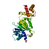
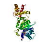
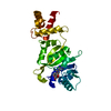
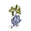
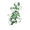
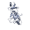
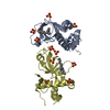
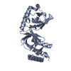
- PDB-1qao: THE STRUCTURE OF THE RRNA METHYLTRANSFERASE ERMC': IMPLICATIONS F... -
+ Open data

- Basic information
| Entry | Database: PDB / ID: 1qao | ||||||
|---|---|---|---|---|---|---|---|
| Title | THE STRUCTURE OF THE RRNA METHYLTRANSFERASE ERMC': IMPLICATIONS FOR THE REACTION MECHANISM | ||||||
 Components Components | ERMC' METHYLTRANSFERASE | ||||||
 Keywords Keywords | TRANSFERASE / BINARY COMPLEX WITH S-ADENOSYLMETHIONINE | ||||||
| Function / homology |  Function and homology information Function and homology information23S rRNA (adenine2085-N6)-dimethyltransferase / 23S rRNA (adenine(2085)-N(6))-dimethyltransferase activity / rRNA (adenine-N6,N6-)-dimethyltransferase activity / response to antibiotic / RNA binding / cytosol Similarity search - Function | ||||||
| Biological species |  | ||||||
| Method |  X-RAY DIFFRACTION / Resolution: 2.7 Å X-RAY DIFFRACTION / Resolution: 2.7 Å | ||||||
 Authors Authors | Schluckebier, G. / Zhong, P. / Stewart, K.D. / Kavanaugh, T.J. / Abad-Zapatero, C. | ||||||
 Citation Citation | Journal: J.Mol.Biol. / Year: 1999 Title: The 2.2 A structure of the rRNA methyltransferase ErmC' and its complexes with cofactor and cofactor analogs: implications for the reaction mechanism. Authors: Schluckebier, G. / Zhong, P. / Stewart, K.D. / Kavanaugh, T.J. / Abad-Zapatero, C. #1: Journal: Biochemistry / Year: 1998Title: Crystal structure of ErmC', an RNA methyltransferase which mediates antibiotic resistance in bacteria Authors: Bussiere, D.E. / Muchmore, S.W. / Dealwis, C.G. / Schluckebier, G. / Nienaber, V.L. / Edalji, R.P. / Walter, K.A. / Ladror, U.S. / Holzman, T.F. / Abad-Zapatero, C. | ||||||
| History |
|
- Structure visualization
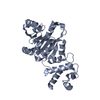
| Structure viewer | Molecule: MolmilJmol/JSmol |
|---|
- Downloads & links
-Download
| PDBx/mmCIF format | 1qao.cif.gz | 60.9 KB | Display | PDBx/mmCIF format |
|---|---|---|---|---|
| PDB format | pdb1qao.ent.gz | 44.7 KB | Display | PDB format |
| PDBx/mmJSON format | 1qao.json.gz | Tree view | PDBx/mmJSON format | |
| Others |  Other downloads Other downloads |
-Validation report
| Arichive directory | https://data.pdbj.org/pub/pdb/validation_reports/qa/1qaoftp://data.pdbj.org/pub/pdb/validation_reports/qa/1qao | HTTPS FTP |
|---|
-Related structure data


-Links
 PDBj
PDBj
- Assembly
| Deposited unit | 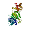
| ||||||||||
|---|---|---|---|---|---|---|---|---|---|---|---|
| 1 |
| ||||||||||
| Unit cell |
|
-Components
| #1: Protein | Mass: 28955.529 Da / Num. of mol.: 1 Source method: isolated from a genetically manipulated source Source: (gene. exp.) |
|---|---|
| #2: Chemical | ChemComp-SAM /   Mass: 398.437 Da / Num. of mol.: 1 / Source method: obtained synthetically / Formula: C15H22N6O5S Mass: 398.437 Da / Num. of mol.: 1 / Source method: obtained synthetically / Formula: C15H22N6O5S |
-Experimental details
-Experiment
| Experiment | Method: X-RAY DIFFRACTION / Number of used crystals: 1 |
|---|
- Sample preparation
| Crystal | Density Matthews: 3.48 Å3/Da / Density % sol: 64.67 % | ||||||||||||||||||||||||||||||||||||||||||||||||||||||
|---|---|---|---|---|---|---|---|---|---|---|---|---|---|---|---|---|---|---|---|---|---|---|---|---|---|---|---|---|---|---|---|---|---|---|---|---|---|---|---|---|---|---|---|---|---|---|---|---|---|---|---|---|---|---|---|
| Crystal grow | Temperature: 298 K / Method: vapor diffusion, hanging drop / pH: 7.8 Details: PEG 8000, ammonium acetate, pH 7.8 at 298 K, VAPOR DIFFUSION, HANGING DROP | ||||||||||||||||||||||||||||||||||||||||||||||||||||||
| Crystal grow | *PLUS pH: 7.5 | ||||||||||||||||||||||||||||||||||||||||||||||||||||||
| Components of the solutions | *PLUS
|
-Data collection
| Diffraction | Mean temperature: 100 K |
|---|---|
| Diffraction source | Source: ROTATING ANODE / Type: RIGAKU RU200 / Wavelength: 1.5418 |
| Detector | Type: RIGAKU RAXIS IIC / Detector: IMAGE PLATE / Date: Jun 11, 1997 |
| Radiation | Protocol: SINGLE WAVELENGTH / Monochromatic (M) / Laue (L): M / Scattering type: x-ray |
| Radiation wavelength | Wavelength: 1.5418 Å / Relative weight: 1 |
| Reflection | Resolution: 2.7→50 Å / Num. all: 11313 / Num. obs: 11313 / % possible obs: 95.5 % / Observed criterion σ(F): 0 / Observed criterion σ(I): 0 / Redundancy: 3.2 % / Biso Wilson estimate: 68 Å2 / Rmerge(I) obs: 0.039 / Net I/σ(I): 15.1 |
| Reflection shell | Resolution: 2.7→2.8 Å / Redundancy: 3.2 % / Rmerge(I) obs: 0.298 / Num. unique all: 1113 / % possible all: 96.6 |
| Reflection | *PLUS Num. measured all: 36172 |
| Reflection shell | *PLUS % possible obs: 96.6 % / Mean I/σ(I) obs: 4.4 |
- Processing
| Software |
| |||||||||||||||
|---|---|---|---|---|---|---|---|---|---|---|---|---|---|---|---|---|
| Refinement | Resolution: 2.7→50 Å / σ(F): 0 / σ(I): 0 / Stereochemistry target values: Engh & Huber
| |||||||||||||||
| Refinement step | Cycle: LAST / Resolution: 2.7→50 Å
| |||||||||||||||
| Refine LS restraints |
| |||||||||||||||
| Software | *PLUS Name: CNS / Classification: refinement | |||||||||||||||
| Refinement | *PLUS Highest resolution: 2.7 Å / Lowest resolution: 50 Å / σ(F): 0 / Rfactor obs: 0.23 / Rfactor Rwork: 0.23 | |||||||||||||||
| Solvent computation | *PLUS | |||||||||||||||
| Displacement parameters | *PLUS | |||||||||||||||
| Refine LS restraints | *PLUS
|
