 Movie
Movie Controller

[English] 日本語
 Yorodumi
- PDB-1pz1: Structure of NADPH-dependent family 11 aldo-keto reductase AKR11B... -
+ Open data

- Basic information
| Entry | Database: PDB / ID: 1pz1 | ||||||
|---|---|---|---|---|---|---|---|
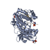
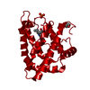
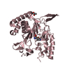
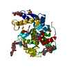
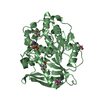
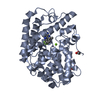
| Title | Structure of NADPH-dependent family 11 aldo-keto reductase AKR11B(holo) | ||||||
 Components Components | General stress protein 69 | ||||||
 Keywords Keywords | OXIDOREDUCTASE / beta-alpha barrel / aldo-keto reductase / TIM barrel | ||||||
| Function / homology |  Function and homology information Function and homology informationOxidoreductases; Acting on the CH-OH group of donors; With NAD+ or NADP+ as acceptor / oxidoreductase activity / cytosol Similarity search - Function | ||||||
| Biological species |  | ||||||
| Method |  X-RAY DIFFRACTION / SYNCHROTRON / MAD / Resolution: 2.2 Å X-RAY DIFFRACTION / SYNCHROTRON / MAD / Resolution: 2.2 Å | ||||||
 Authors Authors | Ehrensberger, A.H. / Wilson, D.K. | ||||||
 Citation Citation | Journal: J.Mol.Biol. / Year: 2004 Title: Structural and Catalytic Diversity in the Two Family 11 Aldo-keto Reductases Authors: Ehrensberger, A.H. / Wilson, D.K. | ||||||
| History |
|
- Structure visualization
| Structure viewer | Molecule: MolmilJmol/JSmol |
|---|
- Downloads & links
-Download
| PDBx/mmCIF format | 1pz1.cif.gz | 153.7 KB | Display | PDBx/mmCIF format |
|---|---|---|---|---|
| PDB format | pdb1pz1.ent.gz | 121.3 KB | Display | PDB format |
| PDBx/mmJSON format | 1pz1.json.gz | Tree view | PDBx/mmJSON format | |
| Others |  Other downloads Other downloads |
-Validation report
| Arichive directory | https://data.pdbj.org/pub/pdb/validation_reports/pz/1pz1ftp://data.pdbj.org/pub/pdb/validation_reports/pz/1pz1 | HTTPS FTP |
|---|
-Related structure data


-Links
 PDBj
PDBj
- Assembly
| Deposited unit | 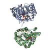
| ||||||||
|---|---|---|---|---|---|---|---|---|---|
| 1 | 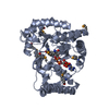
| ||||||||
| 2 | 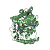
| ||||||||
| Unit cell |
| ||||||||
| Details | The biological unit consists of a monomer. Each asymmetric units consists of two biological units. |
-Components
| #1: Protein | Mass: 37957.617 Da / Num. of mol.: 2 Source method: isolated from a genetically manipulated source Source: (gene. exp.) #2: Chemical |   Mass: 743.405 Da / Num. of mol.: 2 / Source method: obtained synthetically / Formula: C21H28N7O17P3 Mass: 743.405 Da / Num. of mol.: 2 / Source method: obtained synthetically / Formula: C21H28N7O17P3#3: Water | ChemComp-HOH / |  Mass: 18.015 Da / Num. of mol.: 413 / Source method: isolated from a natural source / Formula: H2O Mass: 18.015 Da / Num. of mol.: 413 / Source method: isolated from a natural source / Formula: H2OHas protein modification | Y | |
|---|
-Experimental details
-Experiment
| Experiment | Method: X-RAY DIFFRACTION / Number of used crystals: 1 |
|---|
- Sample preparation
| Crystal | Density Matthews: 2.69 Å3/Da / Density % sol: 54.27 % | ||||||||||||||||||||||||||||||||||||
|---|---|---|---|---|---|---|---|---|---|---|---|---|---|---|---|---|---|---|---|---|---|---|---|---|---|---|---|---|---|---|---|---|---|---|---|---|---|
| Crystal grow | Temperature: 277 K / Method: vapor diffusion, hanging drop / pH: 7.74 Details: PEG 8000, sodium acetate, imidazole, pH 7.74, VAPOR DIFFUSION, HANGING DROP, temperature 277K | ||||||||||||||||||||||||||||||||||||
| Crystal grow | *PLUS Temperature: 4 ℃ / Method: vapor diffusion, hanging drop | ||||||||||||||||||||||||||||||||||||
| Components of the solutions | *PLUS
|
-Data collection
| Diffraction | Mean temperature: 100 K | ||||||||||||
|---|---|---|---|---|---|---|---|---|---|---|---|---|---|
| Diffraction source | Source: SYNCHROTRON / Site: SSRL  / Beamline: BL9-2 / Wavelength: 0.97903, 0.88557, 0.97922 / Beamline: BL9-2 / Wavelength: 0.97903, 0.88557, 0.97922 | ||||||||||||
| Detector | Type: ADSC QUANTUM 4 / Detector: CCD / Date: Jan 21, 2003 / Details: double mirror | ||||||||||||
| Radiation | Monochromator: Double crystal / Protocol: MAD / Monochromatic (M) / Laue (L): M / Scattering type: x-ray | ||||||||||||
| Radiation wavelength |
| ||||||||||||
| Reflection | Resolution: 2.2→100 Å / Num. all: 38524 / Num. obs: 36239 / % possible obs: 94.07 % / Observed criterion σ(F): 0 / Observed criterion σ(I): 2 / Rmerge(I) obs: 0.066 / Net I/σ(I): 15.49 | ||||||||||||
| Reflection shell | Resolution: 2.2→2.28 Å / Rmerge(I) obs: 0.209 / Mean I/σ(I) obs: 5.02 / % possible all: 88.2 | ||||||||||||
| Reflection | *PLUS Lowest resolution: 100 Å / Num. obs: 37796 / % possible obs: 98.4 % / Num. measured all: 134257 | ||||||||||||
| Reflection shell | *PLUS Highest resolution: 2.2 Å / % possible obs: 88.2 % |
- Processing
| Software |
| |||||||||||||||||||||||||
|---|---|---|---|---|---|---|---|---|---|---|---|---|---|---|---|---|---|---|---|---|---|---|---|---|---|---|
| Refinement | Method to determine structure: MAD / Resolution: 2.2→20 Å / Isotropic thermal model: ISOTROPIC / σ(F): 0 / Stereochemistry target values: Engh & Huber
| |||||||||||||||||||||||||
| Refinement step | Cycle: LAST / Resolution: 2.2→20 Å
| |||||||||||||||||||||||||
| Refine LS restraints |
| |||||||||||||||||||||||||
| LS refinement shell | Resolution: 2.2→2.28 Å / % reflection obs: 88.2 % | |||||||||||||||||||||||||
| Refinement | *PLUS Highest resolution: 2.2 Å / Lowest resolution: 30 Å | |||||||||||||||||||||||||
| Solvent computation | *PLUS | |||||||||||||||||||||||||
| Displacement parameters | *PLUS |
