 Movie
Movie Controller

[English] 日本語
 Yorodumi
- PDB-1pyb: Crystal Structure of Aquifex aeolicus Trbp111: a Structure-Specif... -
+ Open data

- Basic information
| Entry | Database: PDB / ID: 1pyb | ||||||
|---|---|---|---|---|---|---|---|
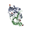
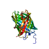
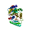
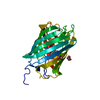
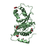
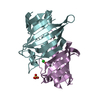
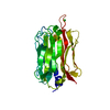
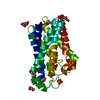
| Title | Crystal Structure of Aquifex aeolicus Trbp111: a Structure-Specific tRNA Binding Protein | ||||||
 Components Components | tRNA-binding protein Trbp111 | ||||||
 Keywords Keywords | RNA BINDING PROTEIN / oligonucleotide / oligosaccharide-binding fold / OB-fold / beta-barrel | ||||||
| Function / homology |  Function and homology information Function and homology informationmethionine-tRNA ligase / methionine-tRNA ligase activity / methionyl-tRNA aminoacylation / tRNA binding / ATP binding / cytoplasm Similarity search - Function | ||||||
| Biological species |   Aquifex aeolicus (bacteria) Aquifex aeolicus (bacteria) | ||||||
| Method |  X-RAY DIFFRACTION / SYNCHROTRON / MOLECULAR REPLACEMENT / Resolution: 2.5 Å X-RAY DIFFRACTION / SYNCHROTRON / MOLECULAR REPLACEMENT / Resolution: 2.5 Å | ||||||
 Authors Authors | Swairjo, M.A. / Morales, A.J. / Wang, C.C. / Ortiz, A.R. / Schimmel, P. | ||||||
 Citation Citation | Journal: Embo J. / Year: 2000 Title: Crystal structure of trbp111: a structure-specific tRNA-binding protein. Authors: Swairjo, M.A. / Morales, A.J. / Wang, C.C. / Ortiz, A.R. / Schimmel, P. #1: Journal: Embo J. / Year: 1999Title: Structure-Specific tRNA-Binding Protein From the Extreme Thermophile Aquifex aeolicus Authors: Morales, A.J. / Swairjo, M.A. / Schimmel, P. | ||||||
| History |
|
- Structure visualization
| Structure viewer | Molecule: MolmilJmol/JSmol |
|---|
- Downloads & links
-Download
| PDBx/mmCIF format | 1pyb.cif.gz | 86.4 KB | Display | PDBx/mmCIF format |
|---|---|---|---|---|
| PDB format | pdb1pyb.ent.gz | 67.8 KB | Display | PDB format |
| PDBx/mmJSON format | 1pyb.json.gz | Tree view | PDBx/mmJSON format | |
| Others |  Other downloads Other downloads |
-Validation report
| Arichive directory | https://data.pdbj.org/pub/pdb/validation_reports/py/1pybftp://data.pdbj.org/pub/pdb/validation_reports/py/1pyb | HTTPS FTP |
|---|
-Related structure data
| Related structure data |  3ersC  1pxf C: citing same article ( S: Starting model for refinement |
|---|---|
| Similar structure data |


-Links
 PDBj
PDBj
- Assembly
| Deposited unit | 
| ||||||||
|---|---|---|---|---|---|---|---|---|---|
| 1 | 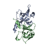
| ||||||||
| 2 | 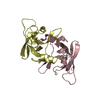
| ||||||||
| Unit cell |
| ||||||||
| Details | The biological unit is the homodimer. There are two homodimers in the asymmetric unit. dimer1: chains A & B, dimer2: chains C & D |
-Components
| #1: Protein | Mass: 12112.133 Da / Num. of mol.: 4 Source method: isolated from a genetically manipulated source Source: (gene. exp.) Aquifex aeolicus (bacteria) / Gene: metG / Plasmid: pTSMg32 / Production host: #2: Water | ChemComp-HOH / |  Mass: 18.015 Da / Num. of mol.: 174 / Source method: isolated from a natural source / Formula: H2O Mass: 18.015 Da / Num. of mol.: 174 / Source method: isolated from a natural source / Formula: H2O |
|---|
-Experimental details
-Experiment
| Experiment | Method: X-RAY DIFFRACTION / Number of used crystals: 1 |
|---|
- Sample preparation
| Crystal | Density Matthews: 3.76 Å3/Da / Density % sol: 67.3 % | ||||||||||||||||||||||||||||||
|---|---|---|---|---|---|---|---|---|---|---|---|---|---|---|---|---|---|---|---|---|---|---|---|---|---|---|---|---|---|---|---|
| Crystal grow | Temperature: 290.15 K / Method: vapor diffusion, hanging drop / pH: 7.2 Details: PEG2000, ammonium sulfate, imidazole, pH 7.2, VAPOR DIFFUSION, HANGING DROP, temperature 290.15K | ||||||||||||||||||||||||||||||
| Crystal grow | *PLUS Temperature: 17 ℃ / Method: vapor diffusion, hanging drop | ||||||||||||||||||||||||||||||
| Components of the solutions | *PLUS
|
-Data collection
| Diffraction | Mean temperature: 100 K |
|---|---|
| Diffraction source | Source: SYNCHROTRON / Site: SSRL  / Beamline: BL7-1 / Wavelength: 1.08 Å / Beamline: BL7-1 / Wavelength: 1.08 Å |
| Detector | Type: MARRESEARCH / Detector: IMAGE PLATE / Date: Aug 3, 1999 |
| Radiation | Monochromator: cylidrically bent single crystals Si(111) with horizontal focus Protocol: SINGLE WAVELENGTH / Monochromatic (M) / Laue (L): M / Scattering type: x-ray |
| Radiation wavelength | Wavelength: 1.08 Å / Relative weight: 1 |
| Reflection | Resolution: 2.4→50 Å / Num. obs: 26611 / % possible obs: 94 % / Observed criterion σ(F): 0 / Observed criterion σ(I): 0 / Redundancy: 2.1 % / Rmerge(I) obs: 0.05 / Rsym value: 0.042 / Net I/σ(I): 9.7 |
| Reflection shell | Resolution: 2.4→2.44 Å / Redundancy: 1.8 % / Rmerge(I) obs: 0.398 / Mean I/σ(I) obs: 2.9 / Num. unique all: 1244 / Rsym value: 0.361 / % possible all: 88.4 |
| Reflection | *PLUS Highest resolution: 2.4 Å / Rmerge(I) obs: 0.042 |
| Reflection shell | *PLUS % possible obs: 88.4 % / Rmerge(I) obs: 0.361 |
- Processing
| Software |
| |||||||||||||||||||||||||
|---|---|---|---|---|---|---|---|---|---|---|---|---|---|---|---|---|---|---|---|---|---|---|---|---|---|---|
| Refinement | Method to determine structure: MOLECULAR REPLACEMENT Starting model: PDB entry 1PXF 1pxf Resolution: 2.5→20 Å / Isotropic thermal model: overall, then group / Cross valid method: THROUGHOUT / σ(F): 0 / σ(I): 0 / Stereochemistry target values: Engh & Huber
| |||||||||||||||||||||||||
| Refinement step | Cycle: LAST / Resolution: 2.5→20 Å
| |||||||||||||||||||||||||
| Refine LS restraints |
| |||||||||||||||||||||||||
| LS refinement shell |
| |||||||||||||||||||||||||
| Xplor file |
| |||||||||||||||||||||||||
| Refinement | *PLUS Lowest resolution: 20 Å / Num. reflection obs: 23601 / % reflection Rfree: 10 % / Rfactor Rfree: 0.22 / Rfactor Rwork: 0.189 | |||||||||||||||||||||||||
| Solvent computation | *PLUS | |||||||||||||||||||||||||
| Displacement parameters | *PLUS |
