 Movie
Movie Controller


+ Open data

- Basic information
| Entry | Database: PDB / ID: 1ps3 | ||||||
|---|---|---|---|---|---|---|---|
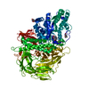
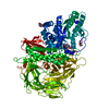
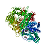
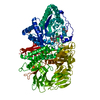
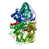
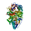
| Title | Golgi alpha-mannosidase II in complex with kifunensine | ||||||
 Components Components | Alpha-mannosidase II | ||||||
 Keywords Keywords | HYDROLASE / glycosyl hydrolase / mannosidase / N-TERMINAL ALPHA-BETA DOMAIN / THREE HELIX BUNDLE / 2 C-TERMINAL BETA BARRELS | ||||||
| Function / homology |  Function and homology information Function and homology informationmannosyl-oligosaccharide 1,3-1,6-alpha-mannosidase / mannosyl-oligosaccharide 1,3-1,6-alpha-mannosidase activity / rhodopsin biosynthetic process / encapsulation of foreign target / Golgi apparatus N-glycan mannose trimming / Reactions specific to the complex N-glycan synthesis pathway / mannosidase activity / alpha-mannosidase activity / mannose metabolic process / N-glycan processing ...mannosyl-oligosaccharide 1,3-1,6-alpha-mannosidase / mannosyl-oligosaccharide 1,3-1,6-alpha-mannosidase activity / rhodopsin biosynthetic process / encapsulation of foreign target / Golgi apparatus N-glycan mannose trimming / Reactions specific to the complex N-glycan synthesis pathway / mannosidase activity / alpha-mannosidase activity / mannose metabolic process / N-glycan processing / Golgi stack / carbohydrate binding / Golgi membrane / endoplasmic reticulum / metal ion binding Similarity search - Function | ||||||
| Biological species |  | ||||||
| Method |  X-RAY DIFFRACTION / SYNCHROTRON / MOLECULAR REPLACEMENT / Resolution: 1.8 Å X-RAY DIFFRACTION / SYNCHROTRON / MOLECULAR REPLACEMENT / Resolution: 1.8 Å | ||||||
 Authors Authors | Shah, N. / Kuntz, D.A. / Rose, D.R. | ||||||
 Citation Citation | Journal: Biochemistry / Year: 2003 Title: Comparison of Kifunensine and 1-Deoxymannojirimycin Binding to Class I and II alpha-Mannosidases Demonstrates Different Saccharide Distortions in Inverting and Retaining Catalytic Mechanisms Authors: Shah, N. / Kuntz, D.A. / Rose, D.R. | ||||||
| History |
|
- Structure visualization
| Structure viewer | Molecule: MolmilJmol/JSmol |
|---|
- Downloads & links
-Download
| PDBx/mmCIF format | 1ps3.cif.gz | 240.3 KB | Display | PDBx/mmCIF format |
|---|---|---|---|---|
| PDB format | pdb1ps3.ent.gz | 187 KB | Display | PDB format |
| PDBx/mmJSON format | 1ps3.json.gz | Tree view | PDBx/mmJSON format | |
| Others |  Other downloads Other downloads |
-Validation report
| Arichive directory | https://data.pdbj.org/pub/pdb/validation_reports/ps/1ps3ftp://data.pdbj.org/pub/pdb/validation_reports/ps/1ps3 | HTTPS FTP |
|---|
-Related structure data
| Related structure data | 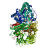 1htyS S: Starting model for refinement |
|---|---|
| Similar structure data |


-Links
 PDBj
PDBj


- Assembly
| Deposited unit | 
| ||||||||
|---|---|---|---|---|---|---|---|---|---|
| 1 |
| ||||||||
| Unit cell |
|
-Components
-Protein / Sugars , 2 types, 2 molecules A
| #1: Protein | Mass: 119593.648 Da / Num. of mol.: 1 / Source method: isolated from a natural source / Source: (natural) References: UniProt: Q24451, mannosyl-oligosaccharide 1,3-1,6-alpha-mannosidase |
|---|---|
| #2: Sugar | ChemComp-NAG /  Type: D-saccharide, beta linking / Mass: 221.208 Da / Num. of mol.: 1 Type: D-saccharide, beta linking / Mass: 221.208 Da / Num. of mol.: 1Source method: isolated from a genetically manipulated source Formula: C8H15NO6 |
-Non-polymers , 4 types, 986 molecules 


| #3: Chemical | ChemComp-ZN /  Mass: 65.409 Da / Num. of mol.: 1 / Source method: obtained synthetically / Formula: Zn Mass: 65.409 Da / Num. of mol.: 1 / Source method: obtained synthetically / Formula: Zn |
|---|---|
| #4: Chemical | ChemComp-KIF /  Mass: 232.191 Da / Num. of mol.: 1 / Source method: obtained synthetically / Formula: C8H12N2O6 Mass: 232.191 Da / Num. of mol.: 1 / Source method: obtained synthetically / Formula: C8H12N2O6 |
| #5: Chemical | ChemComp-MRD / ( Mass: 118.174 Da / Num. of mol.: 1 / Source method: obtained synthetically / Formula: C6H14O2 / Comment: precipitant*YM Mass: 118.174 Da / Num. of mol.: 1 / Source method: obtained synthetically / Formula: C6H14O2 / Comment: precipitant*YM |
| #6: Water | ChemComp-HOH / Mass: 18.015 Da / Num. of mol.: 983 / Source method: isolated from a natural source / Formula: H2O |
-Details
| Has protein modification | Y |
|---|
-Experimental details
-Experiment
| Experiment | Method: X-RAY DIFFRACTION / Number of used crystals: 1 |
|---|
- Sample preparation
| Crystal | Density Matthews: 2.2 Å3/Da / Density % sol: 44.13 % | ||||||||||||||||||||||||
|---|---|---|---|---|---|---|---|---|---|---|---|---|---|---|---|---|---|---|---|---|---|---|---|---|---|
| Crystal grow | Temperature: 298 K / Method: vapor diffusion, hanging drop / pH: 7 Details: PEG 6000, MPD, Tris, pH 7, VAPOR DIFFUSION, HANGING DROP, temperature 298K | ||||||||||||||||||||||||
| Crystal grow | *PLUS Method: vapor diffusion | ||||||||||||||||||||||||
| Components of the solutions | *PLUS
|
-Data collection
| Diffraction | Mean temperature: 100 K |
|---|---|
| Diffraction source | Source: SYNCHROTRON / Site: CHESS  / Beamline: F1 / Wavelength: 0.9504 Å / Beamline: F1 / Wavelength: 0.9504 Å |
| Detector | Type: ADSC QUANTUM 4 / Detector: CCD / Date: Mar 22, 2002 |
| Radiation | Monochromator: Horizontally bent Si(111), assymetrically cut with water cooled Cu block Protocol: SINGLE WAVELENGTH / Monochromatic (M) / Laue (L): M / Scattering type: x-ray |
| Radiation wavelength | Wavelength: 0.9504 Å / Relative weight: 1 |
| Reflection | Resolution: 1.8→30 Å / Num. all: 153548 / Num. obs: 144642 / % possible obs: 85.3 % / Observed criterion σ(F): 3 / Observed criterion σ(I): 2 / Redundancy: 5.25 % / Rmerge(I) obs: 0.072 / Net I/σ(I): 17.2 |
| Reflection shell | Resolution: 1.8→1.83 Å / Rmerge(I) obs: 0.18 / Mean I/σ(I) obs: 4.1 / % possible all: 83.3 |
| Reflection | *PLUS % possible obs: 94.2 % / Num. measured all: 759620 / Rmerge(I) obs: 0.058 |
| Reflection shell | *PLUS % possible obs: 83.3 % / Rmerge(I) obs: 0.18 |
- Processing
| Software |
| ||||||||||||||||||||
|---|---|---|---|---|---|---|---|---|---|---|---|---|---|---|---|---|---|---|---|---|---|
| Refinement | Method to determine structure: MOLECULAR REPLACEMENT Starting model: PDB ENTRY 1HTY Resolution: 1.8→30 Å / Cross valid method: THROUGHOUT / σ(F): 0 / Stereochemistry target values: Engh & Huber
| ||||||||||||||||||||
| Refinement step | Cycle: LAST / Resolution: 1.8→30 Å
| ||||||||||||||||||||
| Refinement | *PLUS Rfactor Rwork: 0.2 | ||||||||||||||||||||
| Solvent computation | *PLUS | ||||||||||||||||||||
| Displacement parameters | *PLUS | ||||||||||||||||||||
| Refine LS restraints | *PLUS
|
