 Movie
Movie Controller

[English] 日本語
 Yorodumi
- PDB-1poi: CRYSTAL STRUCTURE OF GLUTACONATE COENZYME A-TRANSFERASE FROM ACID... -
+ Open data

- Basic information
| Entry | Database: PDB / ID: 1poi | ||||||
|---|---|---|---|---|---|---|---|
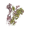
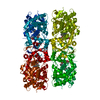
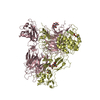
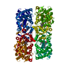
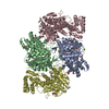
| Title | CRYSTAL STRUCTURE OF GLUTACONATE COENZYME A-TRANSFERASE FROM ACIDAMINOCOCCUS FERMENTANS TO 2.55 ANGSTOMS RESOLUTION | ||||||
 Components Components | (GLUTACONATE COENZYME A-TRANSFERASE) x 2 | ||||||
 Keywords Keywords | TRANSFERASE / COA / GLUTAMATE / PROTEIN FERMENTATION | ||||||
| Function / homology |  Function and homology information Function and homology informationglutaconate CoA-transferase / glutaconate CoA-transferase activity / L-glutamate catabolic process via 2-hydroxyglutarate / cytoplasm Similarity search - Function | ||||||
| Biological species |  Acidaminococcus fermentans (bacteria) Acidaminococcus fermentans (bacteria) | ||||||
| Method |  X-RAY DIFFRACTION / MIR / Resolution: 2.5 Å X-RAY DIFFRACTION / MIR / Resolution: 2.5 Å | ||||||
 Authors Authors | Jacob, U. / Mack, M. / Clausen, T. / Huber, R. / Buckel, W. / Messerschmidt, A. | ||||||
 Citation Citation | Journal: Structure / Year: 1997 Title: Glutaconate CoA-transferase from Acidaminococcus fermentans: the crystal structure reveals homology with other CoA-transferases. Authors: Jacob, U. / Mack, M. / Clausen, T. / Huber, R. / Buckel, W. / Messerschmidt, A. | ||||||
| History |
|
- Structure visualization
| Structure viewer | Molecule: MolmilJmol/JSmol |
|---|
- Downloads & links
-Download
| PDBx/mmCIF format | 1poi.cif.gz | 238 KB | Display | PDBx/mmCIF format |
|---|---|---|---|---|
| PDB format | pdb1poi.ent.gz | 191.1 KB | Display | PDB format |
| PDBx/mmJSON format | 1poi.json.gz | Tree view | PDBx/mmJSON format | |
| Others |  Other downloads Other downloads |
-Validation report
| Summary document | 1poi_validation.pdf.gz | 391 KB | Display | wwPDB validaton report |
|---|---|---|---|---|
| Full document | 1poi_full_validation.pdf.gz | 417.3 KB | Display | |
| Data in XML | 1poi_validation.xml.gz | 25.7 KB | Display | |
| Data in CIF | 1poi_validation.cif.gz | 41.6 KB | Display | |
| Arichive directory | https://data.pdbj.org/pub/pdb/validation_reports/po/1poiftp://data.pdbj.org/pub/pdb/validation_reports/po/1poi | HTTPS FTP |
-Related structure data
| Similar structure data |
|---|


-Links
 PDBj
PDBj- Assembly
| Deposited unit | 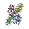
| ||||||||||||
|---|---|---|---|---|---|---|---|---|---|---|---|---|---|
| 1 | 
| ||||||||||||
| Unit cell |
| ||||||||||||
| Components on special symmetry positions |
| ||||||||||||
| Noncrystallographic symmetry (NCS) | NCS oper:
|
-Components
| #1: Protein | Mass: 35390.273 Da / Num. of mol.: 2 Source method: isolated from a genetically manipulated source Source: (gene. exp.) Acidaminococcus fermentans (bacteria) / Cellular location (production host): CYTOSOL / Production host: #2: Protein | Mass: 28566.547 Da / Num. of mol.: 2 Source method: isolated from a genetically manipulated source Source: (gene. exp.) Acidaminococcus fermentans (bacteria) / Cellular location (production host): CYTOSOL / Production host: #3: Chemical | ChemComp-CU / |   Mass: 63.546 Da / Num. of mol.: 1 / Source method: obtained synthetically / Formula: Cu Mass: 63.546 Da / Num. of mol.: 1 / Source method: obtained synthetically / Formula: Cu#4: Water | ChemComp-HOH / |  Mass: 18.015 Da / Num. of mol.: 438 / Source method: isolated from a natural source / Formula: H2O Mass: 18.015 Da / Num. of mol.: 438 / Source method: isolated from a natural source / Formula: H2O |
|---|
-Experimental details
-Experiment
| Experiment | Method: X-RAY DIFFRACTION / Number of used crystals: 2 |
|---|
- Sample preparation
| Crystal | Density Matthews: 2.8 Å3/Da / Density % sol: 56 % | ||||||||||||||||||||||||||||||||||||||||||||||||||||||||||||
|---|---|---|---|---|---|---|---|---|---|---|---|---|---|---|---|---|---|---|---|---|---|---|---|---|---|---|---|---|---|---|---|---|---|---|---|---|---|---|---|---|---|---|---|---|---|---|---|---|---|---|---|---|---|---|---|---|---|---|---|---|---|
| Crystal grow | pH: 5.5 / Details: AMMONIUM SULFATE/PEG PH=5.5 | ||||||||||||||||||||||||||||||||||||||||||||||||||||||||||||
| Crystal grow | *PLUS Temperature: 20 ℃ / Method: vapor diffusion, sitting drop | ||||||||||||||||||||||||||||||||||||||||||||||||||||||||||||
| Components of the solutions | *PLUS
|
-Data collection
| Diffraction | Mean temperature: 298 K |
|---|---|
| Diffraction source | Wavelength: 1.5418 |
| Detector | Type: MARRESEARCH / Detector: IMAGE PLATE / Date: May 1, 1995 |
| Radiation | Monochromatic (M) / Laue (L): M / Scattering type: x-ray |
| Radiation wavelength | Wavelength: 1.5418 Å / Relative weight: 1 |
| Reflection | Resolution: 2.5→7 Å / Num. obs: 37201 / % possible obs: 83 % / Observed criterion σ(I): 0 / Redundancy: 2.8 % / Rmerge(I) obs: 0.098 |
| Reflection shell | Resolution: 2.55→2.64 Å / % possible all: 32 |
- Processing
| Software |
| ||||||||||||||||||||||||||||||||||||||||||||||||||||||||||||
|---|---|---|---|---|---|---|---|---|---|---|---|---|---|---|---|---|---|---|---|---|---|---|---|---|---|---|---|---|---|---|---|---|---|---|---|---|---|---|---|---|---|---|---|---|---|---|---|---|---|---|---|---|---|---|---|---|---|---|---|---|---|
| Refinement | Method to determine structure: MIR / Resolution: 2.5→7 Å / Rfactor Rwork: 0.19 / Rfactor obs: 0.19 / Cross valid method: FREE-R / σ(F): 0 | ||||||||||||||||||||||||||||||||||||||||||||||||||||||||||||
| Displacement parameters | Biso mean: 24 Å2 | ||||||||||||||||||||||||||||||||||||||||||||||||||||||||||||
| Refine analyze | Luzzati coordinate error obs: 0.31 Å | ||||||||||||||||||||||||||||||||||||||||||||||||||||||||||||
| Refinement step | Cycle: LAST / Resolution: 2.5→7 Å
| ||||||||||||||||||||||||||||||||||||||||||||||||||||||||||||
| Refine LS restraints |
| ||||||||||||||||||||||||||||||||||||||||||||||||||||||||||||
| Software | *PLUS Name: X-PLOR / Version: 3.1 / Classification: refinement | ||||||||||||||||||||||||||||||||||||||||||||||||||||||||||||
| Refinement | *PLUS Num. reflection obs: 37201 / Rfactor Rfree: 0.27 | ||||||||||||||||||||||||||||||||||||||||||||||||||||||||||||
| Solvent computation | *PLUS | ||||||||||||||||||||||||||||||||||||||||||||||||||||||||||||
| Displacement parameters | *PLUS |
