[English] 日本語
Yorodumi
- PDB-1pcx: Crystal structure of the COPII coat subunit, Sec24, complexed wit... -

+
Open data


ID or keywords:

Loading...

-
Basic information

Entry
Database: PDB / ID: 1pcx
Title
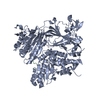
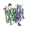
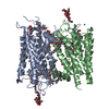
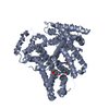
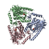
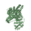
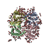
Crystal structure of the COPII coat subunit, Sec24, complexed with a peptide from the SNARE protein Bet1
Components
  • COPII-binding peptide of the protein transport protein BET1
  • Protein transport protein Sec24Protein targeting
KeywordsTRANSPORT PROTEIN
Function / homology
Function and homology information


: / Antigen Presentation: Folding, assembly and peptide loading of class I MHC / Cargo concentration in the ER / Intra-Golgi traffic / positive regulation of ER to Golgi vesicle-mediated transport / COPI-mediated anterograde transport / COPII-mediated vesicle transport / COPII-coated vesicle cargo loading / vesicle fusion / COPII vesicle coat ...: / Antigen Presentation: Folding, assembly and peptide loading of class I MHC / Cargo concentration in the ER / Intra-Golgi traffic / positive regulation of ER to Golgi vesicle-mediated transport / COPI-mediated anterograde transport / COPII-mediated vesicle transport / COPII-coated vesicle cargo loading / vesicle fusion / COPII vesicle coat / SNARE complex / SNAP receptor activity / positive regulation of protein exit from endoplasmic reticulum / signal sequence binding / retrograde vesicle-mediated transport, Golgi to endoplasmic reticulum / fungal-type vacuole membrane / COPII-coated ER to Golgi transport vesicle / endoplasmic reticulum to Golgi vesicle-mediated transport / endoplasmic reticulum exit site / SNARE binding / macroautophagy / intracellular protein transport / protein transport / membrane => GO:0016020 / Golgi membrane / endoplasmic reticulum membrane / endoplasmic reticulum / zinc ion binding
Similarity search - Function
BET1-like protein / BET1, SNARE domain / Sec23/Sec24 helical domain / beta-sandwich domain of Sec23/24 / Zn-finger domain of Sec23/24 / Sec24-like, trunk domain / Zinc finger, Sec23/Sec24-type / Sec23/Sec24, trunk domain / Sec23/Sec24, helical domain / Sec23/Sec24 beta-sandwich ...BET1-like protein / BET1, SNARE domain / Sec23/Sec24 helical domain / beta-sandwich domain of Sec23/24 / Zn-finger domain of Sec23/24 / Sec24-like, trunk domain / Zinc finger, Sec23/Sec24-type / Sec23/Sec24, trunk domain / Sec23/Sec24, helical domain / Sec23/Sec24 beta-sandwich / Zinc finger, Sec23/Sec24-type superfamily / Sec23/Sec24 helical domain superfamily / Sec23/Sec24 zinc finger / Sec23/Sec24 trunk domain / Sec23/Sec24 helical domain / Sec23/Sec24 beta-sandwich domain / Gelsolin-like domain superfamily / Helical region found in SNAREs / t-SNARE coiled-coil homology domain profile. / Target SNARE coiled-coil homology domain / Severin / Severin / Gelsolin-like domain / Gelsolin repeat / von Willebrand factor, type A domain / ADF-H/Gelsolin-like domain superfamily / von Willebrand factor A-like domain superfamily / Four Helix Bundle (Hemerythrin (Met), subunit A) / SH3 type barrels. / Roll / Up-down Bundle / Immunoglobulin-like / Sandwich / Rossmann fold / 3-Layer(aba) Sandwich / Mainly Beta / Mainly Alpha / Alpha Beta
Similarity search - Domain/homology
Protein transport protein BET1 / Protein transport protein SEC24
Similarity search - Component
Biological speciesSaccharomyces cerevisiae (brewer's yeast)
MethodX-RAY DIFFRACTION / SYNCHROTRON / MOLECULAR REPLACEMENT / Resolution: 2.5 Å
AuthorsMossessova, E. / Bickford, L.C. / Goldberg, J.
CitationJournal: Cell(Cambridge,Mass.) / Year: 2003
Title: SNARE selectivity of the COPII coat.
Authors: Mossessova, E. / Bickford, L.C. / Goldberg, J.
History
DepositionMay 18, 2003Deposition site: RCSB / Processing site: RCSB
Revision 1.0Aug 19, 2003Provider: repository / Type: Initial release
Revision 1.1Apr 29, 2008Group: Version format compliance
Revision 1.2Jul 13, 2011Group: Version format compliance
Revision 1.3Feb 14, 2024Group: Data collection / Database references / Derived calculations
Category: chem_comp_atom / chem_comp_bond ...chem_comp_atom / chem_comp_bond / database_2 / struct_conn / struct_site
Item: _database_2.pdbx_DOI / _database_2.pdbx_database_accession ..._database_2.pdbx_DOI / _database_2.pdbx_database_accession / _struct_conn.ptnr1_auth_comp_id / _struct_conn.ptnr1_auth_seq_id / _struct_conn.ptnr1_label_asym_id / _struct_conn.ptnr1_label_atom_id / _struct_conn.ptnr1_label_comp_id / _struct_conn.ptnr1_label_seq_id / _struct_conn.ptnr2_auth_comp_id / _struct_conn.ptnr2_auth_seq_id / _struct_conn.ptnr2_label_asym_id / _struct_conn.ptnr2_label_atom_id / _struct_conn.ptnr2_label_comp_id / _struct_conn.ptnr2_label_seq_id / _struct_site.pdbx_auth_asym_id / _struct_site.pdbx_auth_comp_id / _struct_site.pdbx_auth_seq_id

-
Structure visualization

Structure viewerMolecule:
MolmilJmol/JSmol

Downloads & links

-
Assembly

Deposited unit
A: Protein transport protein Sec24
B: COPII-binding peptide of the protein transport protein BET1
hetero molecules


Theoretical massNumber of molelcules
Total (without water)91,7413
Polymers91,6762
Non-polymers651
Water2,954164
1


  • Idetical with deposited unit
  • defined by author&software
TypeNameSymmetry operationNumber
identity operation1_555x,y,z1
Buried area1000 Å2
ΔGint-3 kcal/mol
Surface area30730 Å2
MethodPISA
Unit cell
Length a, b, c (Å)94.930, 94.930, 198.574
Angle α, β, γ (deg.)90.00, 90.00, 120.00
Int Tables number154
Space group name H-MP3221

-
Components

#1: Protein Protein transport protein Sec24 / Protein targeting / Abnormal nuclear morphology 1


Mass: 90841.672 Da / Num. of mol.: 1 / Source method: isolated from a natural source / Details: Sec24 fragment lacking n-terminal residues 1-116 / Source: (natural) Saccharomyces cerevisiae (brewer's yeast) / References: UniProt: P40482
#2: Protein/peptide COPII-binding peptide of the protein transport protein BET1


Mass: 833.885 Da / Num. of mol.: 1 / Source method: obtained synthetically
Details: THE PEPTIDE WAS CHEMICALLY SYNTHESIZED. THE SEQUENCE OF THE PEPTIDE IS NATURALLY FOUND IN SACCHAROMYCES CEREVISIAE
References: UniProt: P22804
#3: Chemical ChemComp-ZN / ZINC ION


Mass: 65.409 Da / Num. of mol.: 1 / Source method: obtained synthetically / Formula: Zn
#4: Water ChemComp-HOH / water / Water


Mass: 18.015 Da / Num. of mol.: 164 / Source method: isolated from a natural source / Formula: H2O

-
Experimental details

-
Experiment

ExperimentMethod: X-RAY DIFFRACTION / Number of used crystals: 1

-
Sample preparation

CrystalDensity Matthews: 2.82 Å3/Da / Density % sol: 56.34 %
Crystal growTemperature: 298 K / Method: vapor diffusion, hanging drop / pH: 5.8
Details: sodium acetate 1,6-hexanediol, cobalt chloride, pH 5.8, VAPOR DIFFUSION, HANGING DROP, temperature 298K
Crystal grow
*PLUS
Temperature: 4 ℃ / pH: 7.3 / Method: vapor diffusion, hanging drop
Components of the solutions
*PLUS
IDConc.Common nameCrystal-IDSol-IDChemical formulaDetails
150 mg/mlprotein1drop
2200 mM1dropNaCl
320 mMHEPES1droppH7.3
44 mMdithiothreitol1drop
5100 mM1reservoirNaOAcpH5.8
60.35 M1,6-hexanediol1reservoir
74 mM1reservoirCoCl2

-
Data collection

DiffractionMean temperature: 100 K
Diffraction sourceSource: SYNCHROTRON / Site: NSLS / Beamline: X25 / Wavelength: 1 Å
DetectorType: ADSC QUANTUM 4 / Detector: CCD / Date: Sep 12, 2002
RadiationProtocol: SINGLE WAVELENGTH / Monochromatic (M) / Laue (L): M / Scattering type: x-ray
Radiation wavelengthWavelength: 1 Å / Relative weight: 1
ReflectionResolution: 2.5→20 Å / Num. obs: 34136 / % possible obs: 93.8 % / Observed criterion σ(F): 0 / Observed criterion σ(I): 0 / Biso Wilson estimate: 43.6 Å2 / Rmerge(I) obs: 0.044 / Rsym value: 0.0999 / Net I/σ(I): 47.3
Reflection shellResolution: 2.5→2.59 Å / Rmerge(I) obs: 0.3 / Mean I/σ(I) obs: 3.1 / Num. unique all: 3076 / Rsym value: 0.0999 / % possible all: 86.3
Reflection
*PLUS
Num. measured all: 294648
Reflection shell
*PLUS
Highest resolution: 2.5 Å / % possible obs: 86.3 % / Num. unique obs: 3076 / Num. measured obs: 19905 / Rmerge(I) obs: 0.3

-
Processing

Software
NameVersionClassification
CNS1refinement
DENZOdata reduction
SCALEPACKdata scaling
AMoREphasing
RefinementMethod to determine structure: MOLECULAR REPLACEMENT / Resolution: 2.5→19.78 Å / Rfactor Rfree error: 0.007 / Isotropic thermal model: RESTRAINED / Cross valid method: THROUGHOUT / σ(F): 1 / Stereochemistry target values: Engh & Huber
RfactorNum. reflection% reflectionSelection details
Rfree0.259 1560 5 %RANDOM
Rwork0.217 ---
obs0.217 30945 84.6 %-
Solvent computationSolvent model: FLAT MODEL / Bsol: 36.1311 Å2 / ksol: 0.291041 e/Å3
Displacement parametersBiso mean: 71.5 Å2
Baniso -1Baniso -2Baniso -3
1-15.87 Å212.56 Å20 Å2
2--15.87 Å20 Å2
3----31.74 Å2
Refine analyze
FreeObs
Luzzati coordinate error0.44 Å0.36 Å
Luzzati d res low-5 Å
Luzzati sigma a0.53 Å0.51 Å
Refinement stepCycle: LAST / Resolution: 2.5→19.78 Å
ProteinNucleic acidLigandSolventTotal
Num. atoms5951 0 1 164 6116
Refine LS restraints
Refine-IDTypeDev idealDev ideal target
X-RAY DIFFRACTIONc_bond_d0.007
X-RAY DIFFRACTIONc_angle_deg1.3
X-RAY DIFFRACTIONc_dihedral_angle_d23.9
X-RAY DIFFRACTIONc_improper_angle_d0.93
X-RAY DIFFRACTIONc_mcbond_it1.421.5
X-RAY DIFFRACTIONc_mcangle_it2.452
X-RAY DIFFRACTIONc_scbond_it1.872
X-RAY DIFFRACTIONc_scangle_it2.822.5
LS refinement shellResolution: 2.5→2.66 Å / Rfactor Rfree error: 0.029 / Total num. of bins used: 6
RfactorNum. reflection% reflection
Rfree0.389 175 4.4 %
Rwork0.363 3844 -
obs--86.3 %
Xplor file
Refine-IDSerial noParam fileTopol file
X-RAY DIFFRACTION1PROTEIN_REP.PARAMPROTEIN.TOP
X-RAY DIFFRACTION2WATER_REP.PARAMWATER.TOP
X-RAY DIFFRACTION3ION.PARAMION.TOP
X-RAY DIFFRACTION4&_1_PARAMETER_INFILE_4&_1_TOPOLOGY_INFILE_4
X-RAY DIFFRACTION5&_1_PARAMETER_INFILE_5&_1_TOPOLOGY_INFILE_5
Refinement
*PLUS
Highest resolution: 2.5 Å / Lowest resolution: 20 Å / % reflection Rfree: 5 % / Rfactor Rfree: 0.26 / Rfactor Rwork: 0.216
Solvent computation
*PLUS
Displacement parameters
*PLUS
Refine LS restraints
*PLUS
Refine-IDTypeDev ideal
X-RAY DIFFRACTIONc_dihedral_angle_d
X-RAY DIFFRACTIONc_dihedral_angle_deg23.9
X-RAY DIFFRACTIONc_improper_angle_d
X-RAY DIFFRACTIONc_improper_angle_deg0.93

+
About Yorodumi

-
News

-
Feb 9, 2022. New format data for meta-information of EMDB entries

New format data for meta-information of EMDB entries

  • Version 3 of the EMDB header file is now the official format.
  • The previous official version 1.9 will be removed from the archive.

Related info.:EMDB header

External links:wwPDB to switch to version 3 of the EMDB data model

-
Aug 12, 2020. Covid-19 info

Covid-19 info

URL: https://pdbj.org/emnavi/covid19.php

New page: Covid-19 featured information page in EM Navigator.

Related info.:Covid-19 info / Mar 5, 2020. Novel coronavirus structure data

+
Mar 5, 2020. Novel coronavirus structure data

Novel coronavirus structure data

Related info.:Yorodumi Speices / Aug 12, 2020. Covid-19 info

External links:COVID-19 featured content - PDBj / Molecule of the Month (242):Coronavirus Proteases

+
Jan 31, 2019. EMDB accession codes are about to change! (news from PDBe EMDB page)

EMDB accession codes are about to change! (news from PDBe EMDB page)

  • The allocation of 4 digits for EMDB accession codes will soon come to an end. Whilst these codes will remain in use, new EMDB accession codes will include an additional digit and will expand incrementally as the available range of codes is exhausted. The current 4-digit format prefixed with “EMD-” (i.e. EMD-XXXX) will advance to a 5-digit format (i.e. EMD-XXXXX), and so on. It is currently estimated that the 4-digit codes will be depleted around Spring 2019, at which point the 5-digit format will come into force.
  • The EM Navigator/Yorodumi systems omit the EMD- prefix.

Related info.:Q: What is EMD? / ID/Accession-code notation in Yorodumi/EM Navigator

External links:EMDB Accession Codes are Changing Soon! / Contact to PDBj

+
Jul 12, 2017. Major update of PDB

Major update of PDB

  • wwPDB released updated PDB data conforming to the new PDBx/mmCIF dictionary.
  • This is a major update changing the version number from 4 to 5, and with Remediation, in which all the entries are updated.
  • In this update, many items about electron microscopy experimental information are reorganized (e.g. em_software).
  • Now, EM Navigator and Yorodumi are based on the updated data.

External links:wwPDB Remediation / Enriched Model Files Conforming to OneDep Data Standards Now Available in the PDB FTP Archive

-
Yorodumi

Thousand views of thousand structures

  • Yorodumi is a browser for structure data from EMDB, PDB, SASBDB, etc.
  • This page is also the successor to EM Navigator detail page, and also detail information page/front-end page for Omokage search.
  • The word "yorodu" (or yorozu) is an old Japanese word meaning "ten thousand". "mi" (miru) is to see.

Related info.:EMDB / PDB / SASBDB / Comparison of 3 databanks / Yorodumi Search / Aug 31, 2016. New EM Navigator & Yorodumi / Yorodumi Papers / Jmol/JSmol / Function and homology information / Changes in new EM Navigator and Yorodumi

Read more