 Movie
Movie Controller

[English] 日本語
 Yorodumi
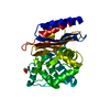
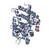
- PDB-1pau: Crystal structure of the complex of apopain with the tetrapeptide... -
+ Open data

- Basic information
| Entry | Database: PDB / ID: 1pau | ||||||
|---|---|---|---|---|---|---|---|
| Title | Crystal structure of the complex of apopain with the tetrapeptide aldehyde inhibitor AC-DEVD-CHO | ||||||
 Components Components |
| ||||||
 Keywords Keywords | HYDROLASE/HYDROLASE INHIBITOR / CYSTEINE PROTEASE / CASPASE-3 / APOPAIN / CPP32 / YAMA / PROTEASE-INHIBITOR COMPLEX / HYDROLASE-HYDROLASE INHIBITOR COMPLEX | ||||||
| Function / homology |  Function and homology information Function and homology informationcaspase-3 / phospholipase A2 activator activity / Stimulation of the cell death response by PAK-2p34 / anterior neural tube closure / intrinsic apoptotic signaling pathway in response to osmotic stress / epithelial cell apoptotic process / leukocyte apoptotic process / glial cell apoptotic process / fibroblast apoptotic process / NADE modulates death signalling ...caspase-3 / phospholipase A2 activator activity / Stimulation of the cell death response by PAK-2p34 / anterior neural tube closure / intrinsic apoptotic signaling pathway in response to osmotic stress / epithelial cell apoptotic process / leukocyte apoptotic process / glial cell apoptotic process / fibroblast apoptotic process / NADE modulates death signalling / positive regulation of pyroptotic inflammatory response / luteolysis / response to cobalt ion / platelet formation / cellular response to staurosporine / cyclin-dependent protein serine/threonine kinase inhibitor activity / response to anesthetic / death-inducing signaling complex / Apoptotic cleavage of cell adhesion proteins / Caspase activation via Dependence Receptors in the absence of ligand / Apoptosis induced DNA fragmentation / SMAC, XIAP-regulated apoptotic response / axonal fasciculation / B cell homeostasis / Activation of caspases through apoptosome-mediated cleavage / Signaling by Hippo / SMAC (DIABLO) binds to IAPs / SMAC(DIABLO)-mediated dissociation of IAP:caspase complexes / death receptor binding / neurotrophin TRK receptor signaling pathway / regulation of synaptic vesicle cycle / negative regulation of cytokine production / execution phase of apoptosis / Other interleukin signaling / negative regulation of B cell proliferation / T cell homeostasis / positive regulation of amyloid-beta formation / Apoptotic cleavage of cellular proteins / negative regulation of activated T cell proliferation / negative regulation of cell cycle / response to amino acid / cell fate commitment / response to glucose / keratinocyte differentiation / response to tumor necrosis factor / Pyroptosis / pyroptotic inflammatory response / response to X-ray / Caspase-mediated cleavage of cytoskeletal proteins / response to UV / response to insulin-like growth factor stimulus / swimming behavior / Degradation of the extracellular matrix / striated muscle cell differentiation / regulation of macroautophagy / protein catabolic process / sensory perception of sound / intrinsic apoptotic signaling pathway / response to glucocorticoid / apoptotic signaling pathway / erythrocyte differentiation / hippocampus development / response to hydrogen peroxide / response to nicotine / protein maturation / regulation of protein stability / protein processing / response to wounding / enzyme activator activity / neuron differentiation / heart development / neuron apoptotic process / response to estradiol / positive regulation of neuron apoptotic process / peptidase activity / response to lipopolysaccharide / protease binding / aspartic-type endopeptidase activity / response to ethanol / response to hypoxia / learning or memory / postsynaptic density / response to xenobiotic stimulus / cysteine-type endopeptidase activity / neuronal cell body / apoptotic process / DNA damage response / protein-containing complex binding / glutamatergic synapse / proteolysis / nucleoplasm / nucleus / cytosol / cytoplasm Similarity search - Function | ||||||
| Biological species |  Homo sapiens (human) Homo sapiens (human) | ||||||
| Method |  X-RAY DIFFRACTION / MOLECULAR REPLACEMENT / Resolution: 2.5 Å X-RAY DIFFRACTION / MOLECULAR REPLACEMENT / Resolution: 2.5 Å | ||||||
 Authors Authors | Rotonda, J. / Becker, J.W. | ||||||
 Citation Citation | Journal: Nat.Struct.Biol. / Year: 1996 Title: The three-dimensional structure of apopain/CPP32, a key mediator of apoptosis. Authors: Rotonda, J. / Nicholson, D.W. / Fazil, K.M. / Gallant, M. / Gareau, Y. / Labelle, M. / Peterson, E.P. / Rasper, D.M. / Ruel, R. / Vaillancourt, J.P. / Thornberry, N.A. / Becker, J.W. #1: Journal: Nature / Year: 1995Title: Identification and Inhibition of the Ice/Ced-3 Protease Necessary for Mammalian Apoptosis Authors: Nicholson, D.W. / Ali, A. / Thornberry, N.A. / Vaillancourt, J.P. / Ding, C.K. / Gallant, M. / Gareau, Y. / Griffin, P.R. / Labelle, M. / Lazebnik, Y.A. / Munday, N.A. / Raju, S.M. / ...Authors: Nicholson, D.W. / Ali, A. / Thornberry, N.A. / Vaillancourt, J.P. / Ding, C.K. / Gallant, M. / Gareau, Y. / Griffin, P.R. / Labelle, M. / Lazebnik, Y.A. / Munday, N.A. / Raju, S.M. / Smulson, M.E. / Yamin, T.T. / Yu, V.L. / Miller, D.K. | ||||||
| History |
|
- Structure visualization
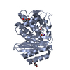
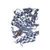
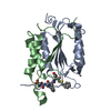
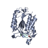
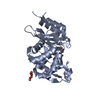
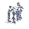
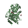
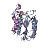
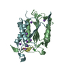
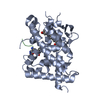
| Structure viewer | Molecule: MolmilJmol/JSmol |
|---|
- Downloads & links
-Download
| PDBx/mmCIF format | 1pau.cif.gz | 55.2 KB | Display | PDBx/mmCIF format |
|---|---|---|---|---|
| PDB format | pdb1pau.ent.gz | 42.5 KB | Display | PDB format |
| PDBx/mmJSON format | 1pau.json.gz | Tree view | PDBx/mmJSON format | |
| Others |  Other downloads Other downloads |
-Validation report
| Arichive directory | https://data.pdbj.org/pub/pdb/validation_reports/pa/1pauftp://data.pdbj.org/pub/pdb/validation_reports/pa/1pau | HTTPS FTP |
|---|
-Related structure data
| Related structure data |  1iceS S: Starting model for refinement |
|---|---|
| Similar structure data |


-Links
 PDBj
PDBj


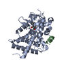
- Assembly
| Deposited unit | 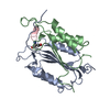
| ||||||||
|---|---|---|---|---|---|---|---|---|---|
| 1 |
| ||||||||
| 2 | 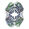
| ||||||||
| 3 | 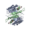
| ||||||||
| Unit cell |
|
-Components
| #1: Protein | Mass: 16639.902 Da / Num. of mol.: 1 Source method: isolated from a genetically manipulated source Source: (gene. exp.) Homo sapiens (human) / Production host:  References: UniProt: P42574, Hydrolases; Acting on peptide bonds (peptidases); Cysteine endopeptidases | ||
|---|---|---|---|
| #2: Protein | Mass: 11910.604 Da / Num. of mol.: 1 Source method: isolated from a genetically manipulated source Source: (gene. exp.) Homo sapiens (human) / Production host: References: UniProt: P42574, Hydrolases; Acting on peptide bonds (peptidases); Cysteine endopeptidases | ||
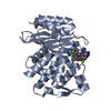
| #3: Protein/peptide | 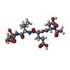  Type: Peptide-like / Class: Inhibitor / Mass: 488.489 Da / Num. of mol.: 1 / Source method: obtained synthetically / References: Ac-Asp-Glu-Val-Asp-Aldehyde Type: Peptide-like / Class: Inhibitor / Mass: 488.489 Da / Num. of mol.: 1 / Source method: obtained synthetically / References: Ac-Asp-Glu-Val-Asp-Aldehyde | ||
| #4: Water | ChemComp-HOH /  Mass: 18.015 Da / Num. of mol.: 34 / Source method: isolated from a natural source / Formula: H2O Mass: 18.015 Da / Num. of mol.: 34 / Source method: isolated from a natural source / Formula: H2O | ||
| Compound details | THE INHIBITOR IS COVALENTLY| Sequence details | AMINO ACID RESIDUES ARE NUMBERED TO FACILITATE COMPARISON WITH THE INTERLEUKIN 1-BETA CONVERTING ...AMINO ACID RESIDUES ARE NUMBERED TO FACILITATE | |
-Experimental details
-Experiment
| Experiment | Method: X-RAY DIFFRACTION / Number of used crystals: 1 |
|---|
- Sample preparation
| Crystal | Density Matthews: 2.47 Å3/Da / Density % sol: 51 % | ||||||||||||||||||||||||||||||||||||||||||||||||||||||
|---|---|---|---|---|---|---|---|---|---|---|---|---|---|---|---|---|---|---|---|---|---|---|---|---|---|---|---|---|---|---|---|---|---|---|---|---|---|---|---|---|---|---|---|---|---|---|---|---|---|---|---|---|---|---|---|
| Crystal grow | Method: vapor diffusion, hanging drop / pH: 5 Details: HANGING DROP VAPOR DIFFUSION. 1.5 MICROLITER DROPS OF PROTEIN:INHIBITOR SOLUTION (8.7 MG/ML IN 10 MILLIMOLAR TRIS-HCL PH 8.5, 10 MILLIMOLAR DTT, 3 MILLIMOLAR SODIUM AZIDE) WERE MIXED WITH AN ...Details: HANGING DROP VAPOR DIFFUSION. 1.5 MICROLITER DROPS OF PROTEIN:INHIBITOR SOLUTION (8.7 MG/ML IN 10 MILLIMOLAR TRIS-HCL PH 8.5, 10 MILLIMOLAR DTT, 3 MILLIMOLAR SODIUM AZIDE) WERE MIXED WITH AN EQUAL VOLUME OF RESERVOIR BUFFER (7% PEG-6000 (W/W), 0.10 MOLAR SODIUM CITRATE PH 5.0, 10 MILLIMOLAR DTT, 3 MILLIMOLAR SODIUM AZIDE) AND INCUBATED AT ROOM TEMPERATURE, vapor diffusion - hanging drop PH range: 5.0-8.0 | ||||||||||||||||||||||||||||||||||||||||||||||||||||||
| Crystal grow | *PLUS pH: 8.5 / Method: vapor diffusion, hanging drop | ||||||||||||||||||||||||||||||||||||||||||||||||||||||
| Components of the solutions | *PLUS
|
-Data collection
| Diffraction | Mean temperature: 293 K |
|---|---|
| Diffraction source | Source: ROTATING ANODE / Type: RIGAKU RUH2R / Wavelength: 1.5418 |
| Detector | Type: SIEMENS / Detector: AREA DETECTOR / Date: Nov 9, 1995 |
| Radiation | Monochromator: GRAPHITE(002) / Monochromatic (M) / Laue (L): M / Scattering type: x-ray |
| Radiation wavelength | Wavelength: 1.5418 Å / Relative weight: 1 |
| Reflection | Resolution: 2.5→36 Å / Num. obs: 8929 / % possible obs: 87.1 % / Observed criterion σ(I): 0 / Redundancy: 2.33 % / Biso Wilson estimate: 31.34 Å2 / Rmerge(I) obs: 0.0634 / Rsym value: 0.0555 / Net I/σ(I): 17.4 |
| Reflection shell | Resolution: 2.5→2.59 Å / Redundancy: 2.27 % / Rmerge(I) obs: 0.257 / Mean I/σ(I) obs: 2.59 / Rsym value: 0.262 / % possible all: 87.1 |
| Reflection | *PLUS Num. measured all: 20801 / Rmerge(I) obs: 0.055 |
| Reflection shell | *PLUS % possible obs: 87.1 % |
- Processing
| Software |
| ||||||||||||||||||||||||||||||||||||||||||||||||||||||||||||
|---|---|---|---|---|---|---|---|---|---|---|---|---|---|---|---|---|---|---|---|---|---|---|---|---|---|---|---|---|---|---|---|---|---|---|---|---|---|---|---|---|---|---|---|---|---|---|---|---|---|---|---|---|---|---|---|---|---|---|---|---|---|
| Refinement | Method to determine structure: MOLECULAR REPLACEMENT Starting model: PROTEIN COMPONENT OF INTERLEUKIN-1BETA CONVERTING ENZYME (PDB ENTRY 1ICE) Resolution: 2.5→20 Å / Cross valid method: FREE-R / σ(F): 2 Details: THERE IS NO ELECTRON DENSITY FOR RESIDUES A 145 - A 149, A 296 - A 297, B 310 - B 319, AND B 402, PRESUMABLY DUE TO DISORDER. MASS SPECTROMETRY INDICATES THAT THESE RESIDUES ARE PRESENT IN ...Details: THERE IS NO ELECTRON DENSITY FOR RESIDUES A 145 - A 149, A 296 - A 297, B 310 - B 319, AND B 402, PRESUMABLY DUE TO DISORDER. MASS SPECTROMETRY INDICATES THAT THESE RESIDUES ARE PRESENT IN THE SPECIES CRYSTALLIZED.
| ||||||||||||||||||||||||||||||||||||||||||||||||||||||||||||
| Displacement parameters | Biso mean: 23.56 Å2 | ||||||||||||||||||||||||||||||||||||||||||||||||||||||||||||
| Refinement step | Cycle: LAST / Resolution: 2.5→20 Å
| ||||||||||||||||||||||||||||||||||||||||||||||||||||||||||||
| Refine LS restraints |
| ||||||||||||||||||||||||||||||||||||||||||||||||||||||||||||
| LS refinement shell | Resolution: 2.5→2.59 Å
| ||||||||||||||||||||||||||||||||||||||||||||||||||||||||||||
| Xplor file |
| ||||||||||||||||||||||||||||||||||||||||||||||||||||||||||||
| Software | *PLUS Name: X-PLOR / Classification: refinement | ||||||||||||||||||||||||||||||||||||||||||||||||||||||||||||
| Refinement | *PLUS | ||||||||||||||||||||||||||||||||||||||||||||||||||||||||||||
| Solvent computation | *PLUS | ||||||||||||||||||||||||||||||||||||||||||||||||||||||||||||
| Displacement parameters | *PLUS | ||||||||||||||||||||||||||||||||||||||||||||||||||||||||||||
| Refine LS restraints | *PLUS
|
