Mass: 18.015 Da / Num. of mol.: 34 / Source method: isolated from a natural source / Formula: H2O
Compound details
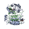
THE DISULFIDE BOND BETWEEN CYS 136 AND CYS 363 REPORTED HERE IS OBSERVED IN SOME OF THE X-RAY DATA ...THE DISULFIDE BOND BETWEEN CYS 136 AND CYS 363 REPORTED HERE IS OBSERVED IN SOME OF THE X-RAY DATA SETS, BUT NOT ALL. THE TWO RESIDUES REMAIN ADJACENT TO ONE ANOTHER IN THOSE INSTANCES WHERE THE SIDE CHAINS ARE NOT POSITIONED TO FORM THE S-S BOND. THERE IS A COVALENT BOND BETWEEN ATOM SG OF CYS285A AND ATOM C OF ASA289T, FORMING A THIOHEMIACETAL
Has protein modification
Y
-
Experimental details
-
Experiment
Experiment
Method: X-RAY DIFFRACTION
-
Sample preparation
Crystal
Density Matthews: 2.86 Å3/Da / Density % sol: 57.03 %
Highest resolution: 2.6 Å / Lowest resolution: 20 Å / % possible obs: 90 % / Rmerge(I) obs: 0.083
-
Processing
Software
Name
Classification
X-PLOR
modelbuilding
X-PLOR
refinement
X-PLOR
phasing
Refinement
Resolution: 2.6→7 Å / σ(F): 1 Details: THE ICE STRUCTURE WAS SOLVED WITH A TETRAPEPTIDE ALDEHYDE REPRESENTED BY CHAIN T COVALENTLY ATTACHED TO THE SULPHUR ATOM OF CYSTEINE 285.
Rfactor
Num. reflection
Rwork
0.19
-
obs
0.19
9123
Refinement step
Cycle: LAST / Resolution: 2.6→7 Å
Protein
Nucleic acid
Ligand
Solvent
Total
Num. atoms
2062
0
0
34
2096
Refine LS restraints
Refine-ID
Type
Dev ideal
X-RAY DIFFRACTION
x_bond_d
0.01
X-RAY DIFFRACTION
x_bond_d_na
X-RAY DIFFRACTION
x_bond_d_prot
X-RAY DIFFRACTION
x_angle_d
X-RAY DIFFRACTION
x_angle_d_na
X-RAY DIFFRACTION
x_angle_d_prot
X-RAY DIFFRACTION
x_angle_deg
2.82
X-RAY DIFFRACTION
x_angle_deg_na
X-RAY DIFFRACTION
x_angle_deg_prot
X-RAY DIFFRACTION
x_dihedral_angle_d
X-RAY DIFFRACTION
x_dihedral_angle_d_na
X-RAY DIFFRACTION
x_dihedral_angle_d_prot
X-RAY DIFFRACTION
x_improper_angle_d
X-RAY DIFFRACTION
x_improper_angle_d_na
X-RAY DIFFRACTION
x_improper_angle_d_prot
X-RAY DIFFRACTION
x_mcbond_it
X-RAY DIFFRACTION
x_mcangle_it
X-RAY DIFFRACTION
x_scbond_it
X-RAY DIFFRACTION
x_scangle_it
Refinement
*PLUS
Rfactor obs: 0.19 / Rfactor Rwork: 0.19
Solvent computation
*PLUS
Displacement parameters
*PLUS
+
About Yorodumi
-
News
-
Feb 9, 2022. New format data for meta-information of EMDB entries
New format data for meta-information of EMDB entries
Version 3 of the EMDB header file is now the official format.
The previous official version 1.9 will be removed from the archive.
In the structure databanks used in Yorodumi, some data are registered as the other names, "COVID-19 virus" and "2019-nCoV". Here are the details of the virus and the list of structure data.
Jan 31, 2019. EMDB accession codes are about to change! (news from PDBe EMDB page)
EMDB accession codes are about to change! (news from PDBe EMDB page)
The allocation of 4 digits for EMDB accession codes will soon come to an end. Whilst these codes will remain in use, new EMDB accession codes will include an additional digit and will expand incrementally as the available range of codes is exhausted. The current 4-digit format prefixed with “EMD-” (i.e. EMD-XXXX) will advance to a 5-digit format (i.e. EMD-XXXXX), and so on. It is currently estimated that the 4-digit codes will be depleted around Spring 2019, at which point the 5-digit format will come into force.
The EM Navigator/Yorodumi systems omit the EMD- prefix.
Related info.:Q: What is EMD? / ID/Accession-code notation in Yorodumi/EM Navigator
Yorodumi is a browser for structure data from EMDB, PDB, SASBDB, etc.
This page is also the successor to EM Navigator detail page, and also detail information page/front-end page for Omokage search.
The word "yorodu" (or yorozu) is an old Japanese word meaning "ten thousand". "mi" (miru) is to see.
Related info.:EMDB / PDB / SASBDB / Comparison of 3 databanks / Yorodumi Search / Aug 31, 2016. New EM Navigator & Yorodumi / Yorodumi Papers / Jmol/JSmol / Function and homology information / Changes in new EM Navigator and Yorodumi
 Movie
Movie Controller


 Open data
 Basic information
 Components
 Keywords
 Function and homology information
 Homo sapiens (human)
 X-RAY DIFFRACTION / Resolution: 2.6 Å
  Authors
 Citation
 Structure visualization
 Downloads & links
 Other downloads
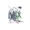


 PDBj
PDBj


 Assembly
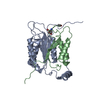


 Type: Peptide-like / Class: Inhibitor / Mass: 476.523 Da / Num. of mol.: 1 / Source method: obtained synthetically / References: Ac-Tyr-Val-Ala-Asp-Aldehyde
 Mass: 18.015 Da / Num. of mol.: 34 / Source method: isolated from a natural source / Formula: H2O
 Sample preparation
 Processing